|  Raymond
V. Damadian, MD, conceived the IDEA
of using NMR (MR) to detect medical disease and proposed
the MR body scanner to accomplish it. To prove its feasibility,
he conducted experiments and DISCOVERED
that cancer tissues produce abnormal NMR signals
compared to normal tissues, with relaxation times that
are markedly elevated relative to normal tissues. He
also DISCOVEREDthat
the healthy tissues themselves exhibit significant differences
in NMR relaxation times*.
The relaxation differences among the normal tissues
supply the contrast needed to see anatomic detail that
was missing in other medical imaging technologies (x-ray
and ultrasound). Recognizing that the abnormal NMR signals
generated by cancers could be used to detect cancers
non-invasively, he went on to build the first whole
body magnetic resonance scanner, which he named Indomitable,
and to achieve the first MRI scan of the live human
body, as well as the first scan of a patient with cancer.
The tissue signals
he DISCOVERED
and their marked differences among the normal tissues
and also between normal tissue and diseased tissue have
remained the source of all MRI images today. Raymond
V. Damadian, MD, conceived the IDEA
of using NMR (MR) to detect medical disease and proposed
the MR body scanner to accomplish it. To prove its feasibility,
he conducted experiments and DISCOVERED
that cancer tissues produce abnormal NMR signals
compared to normal tissues, with relaxation times that
are markedly elevated relative to normal tissues. He
also DISCOVEREDthat
the healthy tissues themselves exhibit significant differences
in NMR relaxation times*.
The relaxation differences among the normal tissues
supply the contrast needed to see anatomic detail that
was missing in other medical imaging technologies (x-ray
and ultrasound). Recognizing that the abnormal NMR signals
generated by cancers could be used to detect cancers
non-invasively, he went on to build the first whole
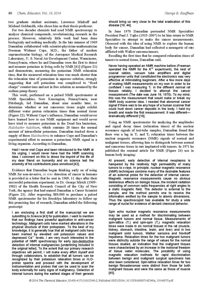
body magnetic resonance scanner, which he named Indomitable,
and to achieve the first MRI scan of the live human
body, as well as the first scan of a patient with cancer.
The tissue signals
he DISCOVERED
and their marked differences among the normal tissues
and also between normal tissue and diseased tissue have
remained the source of all MRI images today.
* The relaxation time differences between cancer tissue and normal tissue and within the normal tissues themselves were the result of differences in the mobility of the water molecules within cancer tissues, relative to their mobility within the normal tissues, and also to the differences in water mobility within the normal tissues themselves. The NMR signal decay time (relaxation time) of the water proton NMR signal is very sensitive to the degree of mobility of the highly mobile water molecules within the examined sample, e.g., the highly mobile water molecules within a sample of liquid water produce a T2 relaxation time of 3,000 milliseconds, while their immobilization in their positions in the crystal lattice of ice produce a T2 relaxation time of .019 milliseconds (a 157,000 times shorter T2 relaxation time for the immobilized water molecules of ice).
The development of the MRI was dependent
on the appearance on the scene in 1969 of some MD, like
Dr. Damadian, experienced BOTH
in the clinical care of patients
and in the technology of NMR.
Up until that point, the NMR had been in use almost
exclusively by chemists to perform test-tube analyses
of chemical samples. The prospect of performing anything
like the scan of a live human body with the existing
23 year old 2¼" NMR test-tube analyzer had
never been imagined. Like any clinically experienced
physician, however, Dr. Damadian was well aware of the
painful experiences eventually endured by almost every
physician during that time period i.e. learning unexpectedly
at autopsy for the first time, of the existence of long
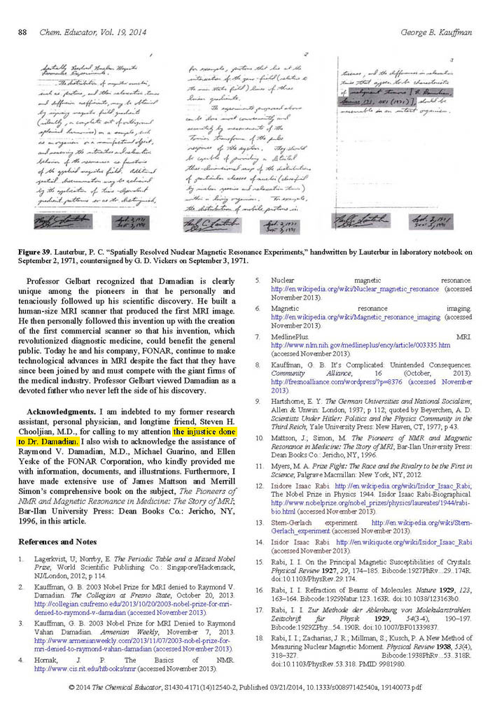
standing and widespread fatal lesions within the body's
vital organ soft tissues (e.g. brain, liver, heart,
intestine, kidney…) that were being poorly visualized
by the imaging technologies of the day (conventional
x-ray) 1 and going UNDETECTED.
Dr. Damadian concluded that much more sensitive DETECTION
of lesions, such as cancer wherever they might have
spread within the human body, would mean much better
monitoring of the effectiveness of the pharmaceutical
regimens being employed. More sensitive DETECTION
would enable the addition of new treatment agents or
further dosage adjustments, if the regimens in use were
proving insufficient. In fact, as it has turned out,
except for the abnormal NMR signals
of diseased tissue DISCOVERED
by Dr. Damadian, FATAL DISEASE like cancer would not
be visible or detectable by any MRI.
1 [The CAT scan was not introduced until April 20, 1972 by J. Ambrose and G. Hounsfield at the Atkinson-Morley Hospital at the 32 Congress British Institute of Radiology in their paper "Computerized Axial Tomography (A new means of demonstrating some of the soft tissue structures of the brain without the use of contrast media)".]
NMR = MRI
MRI IS NMR RENAMED.
Upon the advance of NMR (nuclear magnetic resonance)
technology into its medical applications, as a result
of Dr. Damadian's DISCOVERIES,
the medical community preferred the elimination of the
word nuclear to avoid its radioactive connotations (that
were non-existent) and the radiologic community sought
to have the I added to denote NMR scanning as an imaging
technology.
inventor of the MRI
THE TRUTH OF HISTORY, CAMBRIDGE UNIVERSITY PRESS – THE SAME YEAR AS THE NOBEL PRIZE
 “The
initial concept for the medical application of NMR,
as it was then called, originated
with the discovery by Raymond Damadian in 1971 that certain mouse tumours displayed elevated relaxation times compared
with normal tissues in vitro. This exciting
discovery opened
the door for a complete new way of imaging the human
body where the potential contrast between tissues
and disease was many times greater than that offered
by X-ray technology and ultrasound.” “The
initial concept for the medical application of NMR,
as it was then called, originated
with the discovery by Raymond Damadian in 1971 that certain mouse tumours displayed elevated relaxation times compared
with normal tissues in vitro. This exciting
discovery opened
the door for a complete new way of imaging the human
body where the potential contrast between tissues
and disease was many times greater than that offered
by X-ray technology and ultrasound.”
“So what were NMR researchers doing between the forties and the seventies - that's a long time in cultural and scientific terms. The answer: they were doing chemistry, including Lauterbur, a professor of chemistry at the same institution as Damadian. NMR developed into a laboratory spectroscopic technique capable of examining the molecular structure of compounds, until Damadian's ground-breaking discovery in 1971.”
(MRI from Picture to Proton,
Cambridge University Press, 2003, p.2-4)
inventor of the MRI
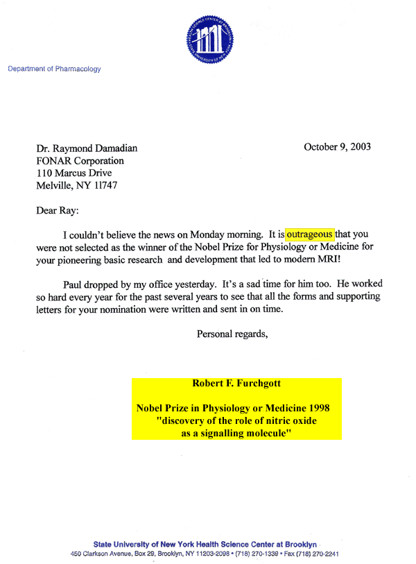
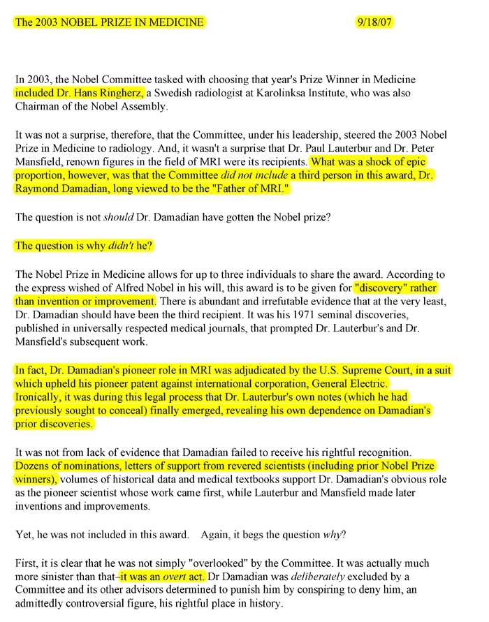
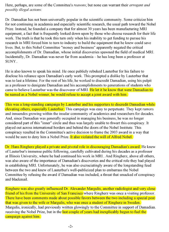
NOBEL VIOLATION OF THE TRUTH
In 2003, The Noble Prize for the MRI
was awarded, not to Dr. Damadian, but to two nuclear
magnetic resonance scientists. One employed a gradient,
invented 50 years earlier by others, to improve the
scanning method discovered
by Dr. Damadian. Another was a member of a group who
found a better way to use gradients to make an MRI image.
Although the prize allowed for three winners, Dr. Damadian
was excluded.
The award is a calculated affront to
the truth of science.
It is also an affront to the WILL of Alfred Nobel, in which he specified that the award in medicine can only be given for “discovery,” not
for technological improvements. (See Fig.19 Nobel Statutes)
Thankfully, the truth of history endures.
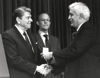 1.TWO PRESIDENTS OF THE UNITED STATES DISAGREE WITH THEM 1.TWO PRESIDENTS OF THE UNITED STATES DISAGREE WITH THEM
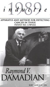
1. President Ronald Reagan awarded the nation's highest honor in technology, The National Medal of Technology to Dr. Damadian and Dr. Lauterbur at the executive Offices of the White House in 1988 "For their independent contributions in conceiving and developing the application of magnetic resonance technology to medical uses including whole-body scanning and diagnostic imaging".
2.President George H.W. Bush inducted Dr. Damadian into The United States National Inventors Hall of Fame.
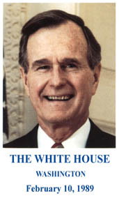 "I am pleased to send warm greetings to everyone present for the 1989 induction ceremony of the National Inventors Hall of Fame. "I am pleased to send warm greetings to everyone present for the 1989 induction ceremony of the National Inventors Hall of Fame.
America must maintain its competitive edge. The challenges before our Nation are too numerous, and the stakes too high, for us to permit the eclipse of that traditional wellspring of our productive genius: our willingness to try new ideas. In the past we have risen to every challenge presented us, and I believe we can rise to the challenges of today. But only if we foster the spirit of invention.
And so I join you in saluting the memory of three great inventors being honored tonight: Westinghouse, Deere, and Langmuir. You are fortunate, I understand, to have a fourth great inventor with you: Dr. Raymond Damadian, whose medical inventions are saving lives around the world. In my association with the wonderful INVENT AMERICA! program, I have seen Dr. Damadian at work, captivating young imaginations with the fires of his own. I would not be surprised to see him joined in the Hall of Fame by some of those promising young minds. All it takes is imagination and encouragement, and he is an ideal source of both. He is living, reassuring proof that the spirit of invention continues to thrive in our great Nation.
Barbara and I join the American people in congratulating Dr. Damadian and in sending our best wishes to all of you."
2.THREE NOBEL LAUREATES DISAGREE WITH THEM
On the Accomplishment of the World's
First MRI Scan of the Live Human Body 7/3/1977
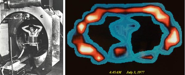
Figure 25
Figure 26
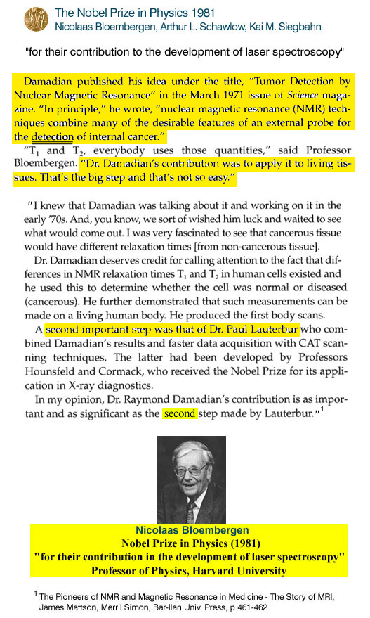
Figure 27
Figue 28
"FRIENDS
OF RAYMOND DAMADIAN"
designate the exclusion of Dr.
Damadian from the 2003 Nobel Prize
A criminal
act WARRANTING A "FULL
CIVIL AND CRIMINAL
INVESTIGATION" !
Professor George Kauffman, Professor
of Chemistry, California State University, upon concluding
his detailed fact finding investigation of the origin
of MRI, entitled "Nobel
Prize for MRI Imaging Denied to Raymond V. Damadian
a Decade Ago", Chemical Educator 2014,
19, 73-90, thanks his colleague Steven H. Chooljian,
M.D. for calling his attention to
"
THE INJUSTICE DONE TO DR. DAMADIAN " (page
88)
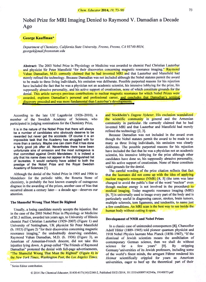
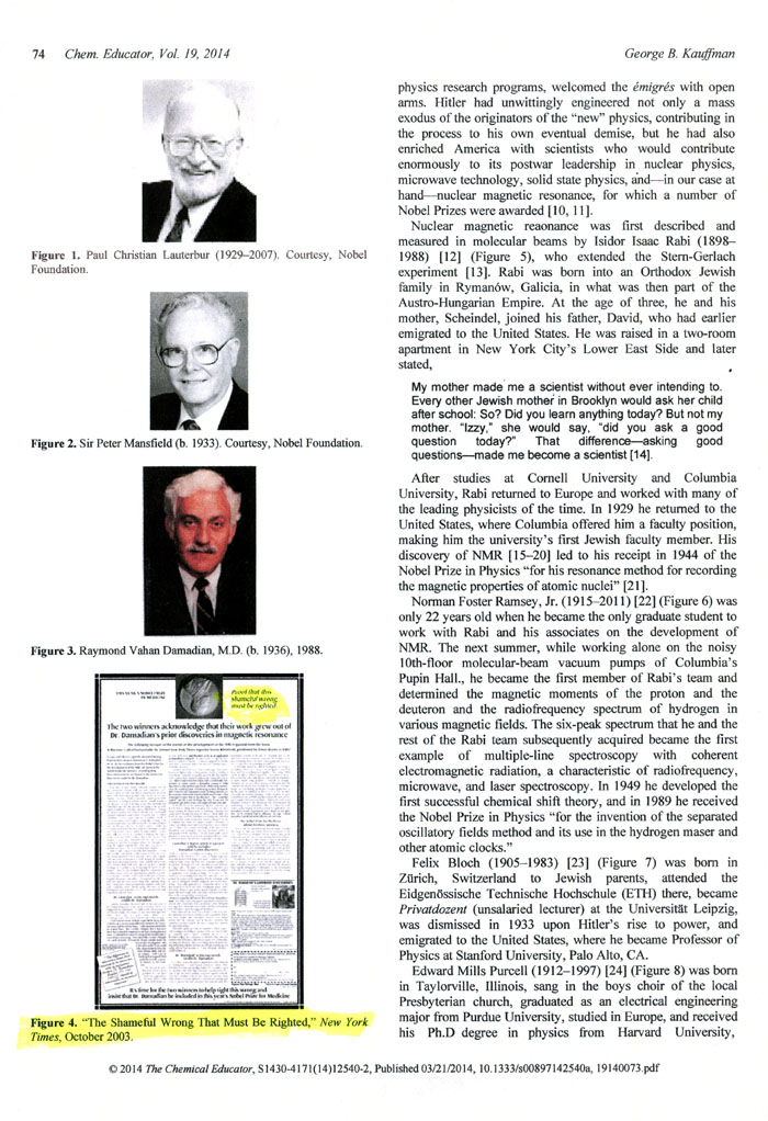
View PDF of the Article: Nobel
Prize for MRI Imaging Denied to Raymond V. Damadian a Decade Ago
The State University of New York
(SUNY)
Downstate Medical Center
expresses its
ANGER
at the 2003 Nobel Committee for their
EXCLUSION
from recognition by the
2003 Nobel Prize Committee in Physiology and Medicine
of SUNY Downstate Medical Center's
role
in providing the faculty, graduate students, medical
students and the building–engineering staff
that achieved the jackhammer reconstruction of Downstate
Medical Center's building to increase the ceiling
height of Dr. Damadian's laboratory and bring to reality
(at Downstate), for
the benefit of mankind,
the first–ever MRI scanner
of the live human body.
"We
are perplexed, disappointed and
angry about the uncomprehensible
exclusion of Professor Raymond Damadian M.D.
from this year's Nobel Prize in Physiology or
Medicine. MRI's entire development rests on
the shoulders of Damadian's discovery
of NMR proton relaxation differences among normal
and diseased tissues and his proposal of external
scanning of NMR relaxation differences in the
human body, published in Science in 1971"
Eugene
Feigelson, M.D.
Dean of the College of Medicine
SUNY Downstate Medical Center
Distinguished Service Professor
Senior Vice President for Biomedical Education
and Research
|
|
|
REMARKABLY
the 2003 Nobel Committee
EXCLUDED
the
ONLY
genuine DISCOVERY*
ENTITLED
under Nobel's statutes for the Nobel Prize in medicine
and
awarded the Nobel Prize in Medicine instead to Lauterbur
and Mansfield for their "INVENTIONS" or
"IMPROVEMENTS" (i.e.
methods) that Nobel specifically EXCLUDED
from the NOBEL PRIZE in Physiology or Medicine.
The
genuine scientific DISCOVERY
that originated MRI was the DISCOVERY
by Dr. Damadian of the tissue NMR signal differences
between diseased and normal tissue that are used to
make the MRI image, the DISCOVERY
of the prolonged relaxation times of diseased
tissues and his new DISCOVERYof
the wide range of NMR signal relaxation times among
the body's vital organ tissues that supply the
PIXEL CONTRAST (IMAGE DETAIL)
that
had been missing from x–ray
technology for the better
part of a century.
His
Genuine new scientific DISCOVERY
provided unprecedented detail in the
visualization of the body's critical vital organs
for the first time
in medical history.
Thus, as Nobel specified, the Nobel Prize in medicine
could be given
ONLY
for DISCOVERY,
i.e. the DISCOVERY
of new scientific phenomena
(e.g. the DISCOVERY
of the change in the NMR signal of the body's tissues
with disease and the DISCOVERY
of the wide variations of the NMR signal relaxations
in
the
body's normal vital organ tissues.
Thus the 2003 Nobel Committee
REMARKABLY EXCLUDED
the
ONLY
GENUINE DISCOVERY
qualified for the Nobel Prize in medicine under the
WILL
of Alfred Nobel and then misrepresented the
contributions of Lauterbur and Mansfield as "discoveries
concerning magnetic resonance imaging" when they
were EXCLUSIVELY METHODS
(and
only methods) ( i.e. the "INVENTIONS"
or "IMPROVEMENTS" EXCLUDED
from Alfred Nobel's statutes for a Nobel prize in
medicine) to implement the
genuine
scientific DISCOVERY
of Dr. Damadian
THAT
MAKES THE IMAGE !
 3.
THE UNITED STATES NATIONAL INVENTOR'S
HALL OF FAME DISAGREES WITH THEM 3.
THE UNITED STATES NATIONAL INVENTOR'S
HALL OF FAME DISAGREES WITH THEM
They inducted Dr. Raymond Damadian in 1989 to join Thomas Edison, Alexander Graham Bell, Samuel Morse, the Wright brothers and the other inventor legends of American history for his invention of "the magnetic resonance imaging (MRI) scanner, which has revolutionized the field of diagnostic medicine".
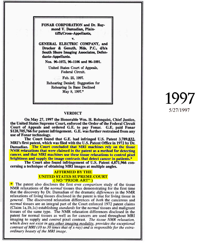
4 . THE UNITED
STATES SUPREME COURT (William Rehnquist, Chief
Justice) AFTER 1.1 MILLION PAGES OF DOCUMENTARY EVIDENCE
DISAGREES WITH THEM.
The First to Propose Scanning the Human Body (1969) by NMR (MRI)
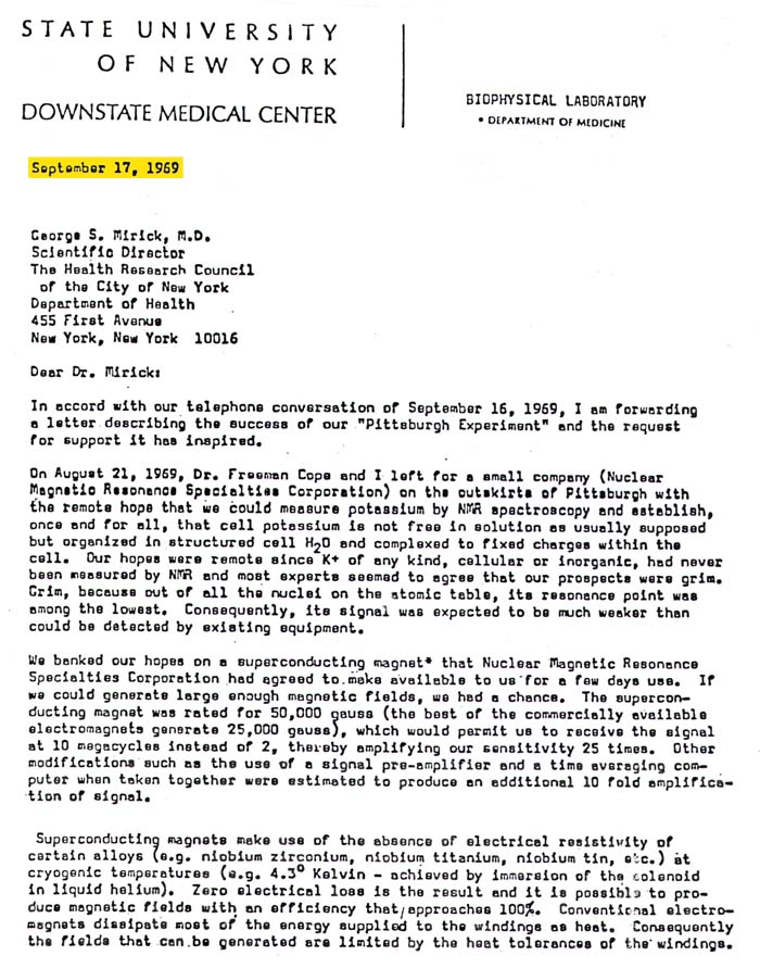
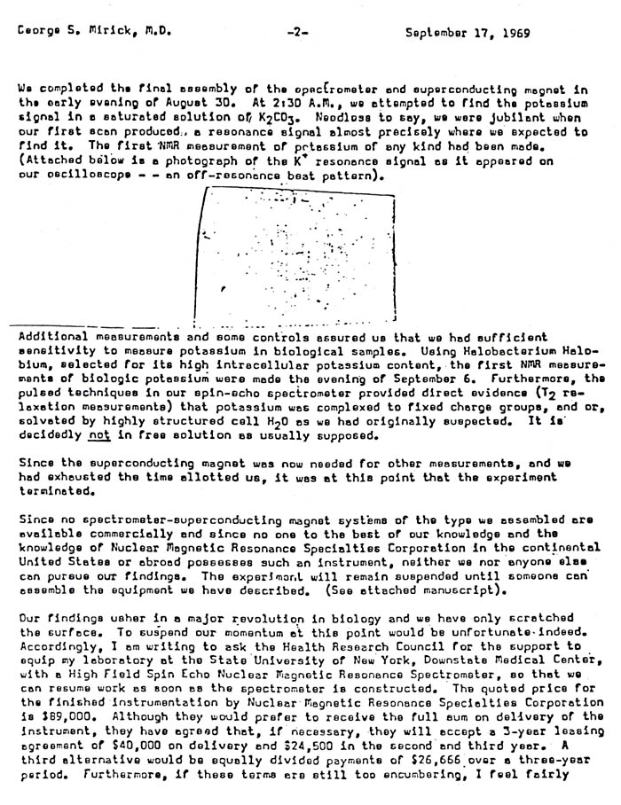
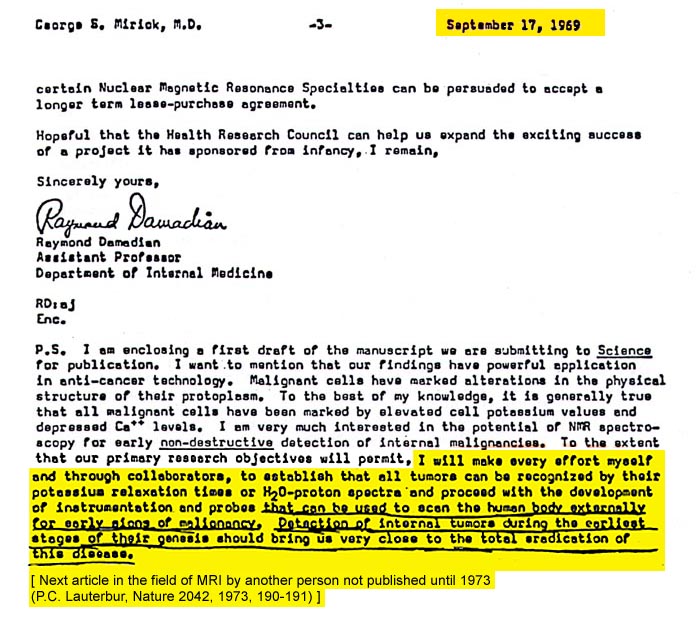
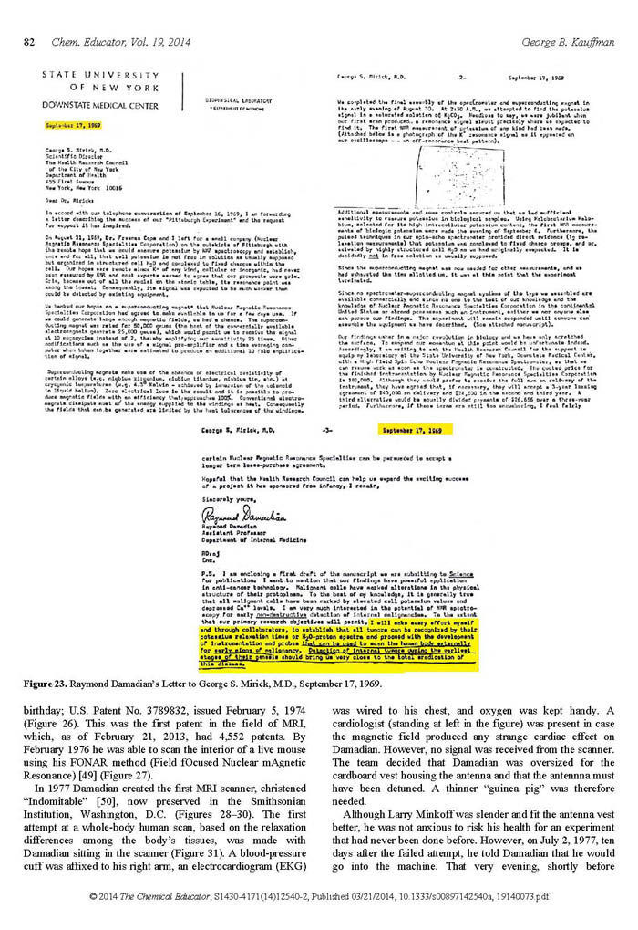
Figure 1.
"I will make every effort myself and through collaborators, to establish that all tumors can be recognized by their potassium relaxation times or H2O-proton spectra and proceed with the DEVELOPMENT OF INSTRUMENTATION and PROBES that can be used to SCAN THE HUMAN BODY EXTERNALLY FOR EARLY SIGNS OF MALIGNANCY. DETECTION
OF INTERNAL TUMORS DURING
THE EARLIEST STAGES OF THEIR GENESIS SHOULD BRING US VERY CLOSE TO THE TOTAL ERADICATION OF THIS DISEASE "
The First to Propose Scanning the Human Body (Spring 1971) by NMR (MRI)
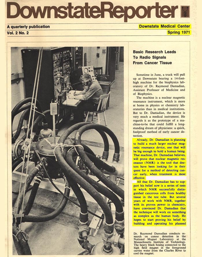
"But several years of work with NMR, together with its proven power in chemistry, have convinced Dr. Damadian that the technique WILL WORK ON SOMETHING AS COMPLEX AS THE HUMAN BODY. HE HOPES TO START PROVING HIS BELIEF BY BUILDING AND OPERATING HIS PLANNED LARGER MACHINE IN THE NEXT TWO YEARS"
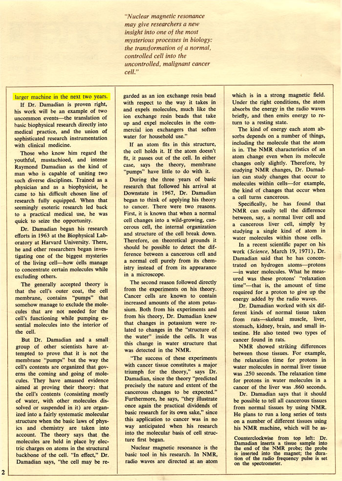
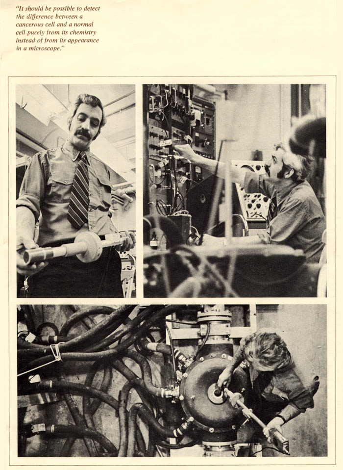
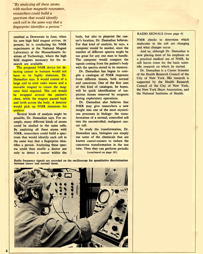
Figure 2.
"The proposed NMR device FOR DETECTING CANCER IN HUMANS would not have to be highly elaborate, Dr. Damadian says. It would consist of a large coil to emit radio waves and a movable magnet to create the magnetic field required. THE COIL WOULD BE WRAPPED AROUND THE PATIENT'S CHEST, WHILE THE MAGNET PASSED BACK AND FORTH ACROSS THE BODY. A DETECTOR WOULD PICK UP NMR EMISSIONS FOR ANALYSIS."
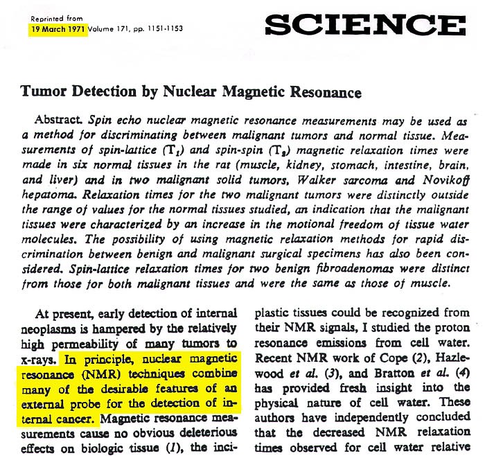
Figure 3.
"At present, EARLY DETECTION of INTERNAL NEOPLASMS is hampered by the relatively high permeability of many tumors to x-rays. In principle, NUCLEAR MAGNETIC RESONANCE (NMR) TECHNIQUES COMBINE MANY OF THE DESIRABLE FEATURES ON AN EXTERNAL PROBE FOR THE DETECTION OF INTERNAL CANCER."
The Signal Makes The
Image !
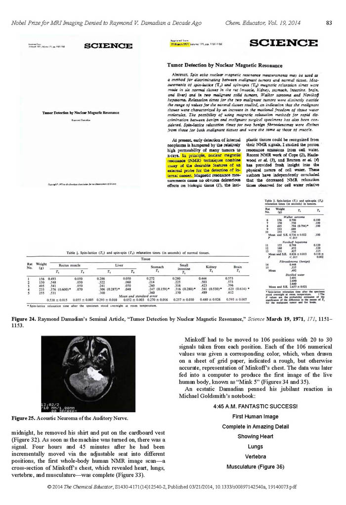
1. R. Damadian, Tumor Detection by Nuclear Magnetic Resonance. Science, 19 March 1971, Vol.171, pp. 1151-1153.
No Signal Differences: 1, NO IMAGE !
Raymond V. Damadian is the medical doctor
who first proposed scanning medical patients by NMR
(nuclear magnetic resonance, the original name of the
MRI) based on his DISCOVERY
of the principle on which all modern MRI is based –
that different tissues emit different NMR signals
in a magnetic field. The amplitude of the signal
determines the brightness of the picture element (pixel)
that the MRI image is composed of.
The Signal

Figure 4a.
The nucleus of the atom possesses a spin. Composed as it is of electrically charged components, protons, its nuclear spin generates a magnetic moment, i.e. the spinning hydrogen nucleus is therefore a two-pole (dipole) magnet with a north magnetic pole and a south magnetic pole. While much smaller, the magnetic fields generated by these spinning positively charged protons are analogous to the magnetic fields generated by their negatively charged counterparts, e.g. the magnetic fields generated by Faraday's induction when electrons spin or move in circular paths such as the magnetic fields generated along the axis of a circular loop of wire as electricity traverses a circular path.
When exposed to a magnetic field, these spinning nuclear magnets, e.g. the hydrogen protons of tissue water (H2O), line up with the magnetic field and quantize, i.e. separate into two populations, a low energy population that magnetically aligns parallel with the applied magnetic field of the MRI magnet and a less populous high energy population that aligns opposite (anti-parallel) to the main magnetic field. The separation into two energy groups, the low energy and the high energy group, generates the prospect of energy transitions between the two energy populations by the application of additional energy, e.g. the application of additional magnetic energy provided by an oscillating energy source such as a radio frequency. Radio Frequency signals (r.f.), are oscillating electro-magnetic fields. In the case of NMR (MR), the magnetic component of the radiofrequency signal is the component that provides the energy necessary to excite some of the low energy nuclear magnet population into
the high energy nuclear population. This nuclear resonance stimulation is achieved in practice with an r.f. transmitter coil that encircles the human body to provide this oscillating magnetic energy in order to excite some of the low energy nuclei (e.g. hydrogen protons of tissue water (H2O)) to transition to the high energy population.
When the transmitter is shut off, the excited (high energy) nuclear spins emit their absorbed radio energy in order to return to their low energy equilibrium (resting) state. The emitted energy is then captured by a radio receiver coil wrapped around the human body. The excitation radiofrequency is tuned to the frequency needed to supply the exact energy (the resonant frequency) necessary to convert the low energy nuclear spins to high energy spins.
As seen in the above Figure
4a (the nuclear signal)
the signal captured
by the receiver coil decays over time (its "relaxation
time") until the original excitation energy that excited
the low energy nuclear spin into the high energy state
is fully dissipated. The time to complete dissipation
of the original excitation energy is called its "relaxation
time". This "relaxation time" varies markedly with the
local anatomic and chemical environment in which the
signal generating
nuclear magnet resides (e.g. the T2 relaxation time
of a water proton in its liquid form is 3,000 mseconds
while the same T2 relaxation time for a water proton
in ice is .019 mseconds). Accordingly, the decay time
(relaxation time) of the water proton NMR signal
is very sensitive to any anatomic changes of tissue
structure. As discovered
by Damadian, the tissue structure changes in the immediate
vicinity of the resonating proton that accompany tissue
disease or the tissue structure variations within the
normal organs themelves (heart muscle, liver, intestine,
etc.— Figure 6) profoundly
affect the relaxation time of this nuclear magnetic
signal.
Tables 1 & 2.
R. Damadian, Tumor Detection by Nuclear
Magnetic Resonance. Science, 19 March 1971, Vol.171,
Tables 1 & 2, pp.
1151-1153. Raymond V. Damadian is the medical doctor
who first proposed scanning medical patients by NMR
(nuclear magnetic resonance, the original name of the
MRI) based on his DISCOVERY
of the principle on which all modern MRI is based —
the different NMR signals
that tissues emit in a magnetic field. The amplitude
of these signals
determines the brightness of the picture elements (pixels)
that the MRI image is composed of. The black rectangle
display is a summary table of the abnormal T1 signal
decay times (relaxation times) of tumor tissue published
in Science [Tables 1 & 2, R. Damadian, Science (1971,
171, p1151) ].
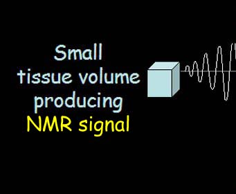
A live NMR signal such as that generated by a small tissue volume connected to an oscilloscope and an audio amplifier so that an example of an NMR signal that generates the MRI image can be directly visualized and heard. [ Click on Sine Wave to Listen ]
THE STRENGTH OF THE SIGNAL1 SETS
THE
PIXEL BRIGHTNESS !
1. The computed strength of the signal (the amplitude) is determined by the signal's decay time (relaxation time). The longer the relaxation time the greater the signal amplitude and the greater the brightness of the picture elements (pixels) that compose the image.
The Signal Makes The Image:
No Signal Differences1, No Image.
1. R. Damadian, Tumor Detection by Nuclear Magnetic
Resonance. Science, 19 March 1971, Vol.171, pp. 1151-1153.
Raymond V. Damadian is the medical doctor who first
proposed scanning medical patients by NMR (nuclear magnetic
resonance, the original name of the MRI) based on his
DISCOVERY
of the principle on which all modern MRI is based —
the different NMR signals
that tissues emit in a magnetic field. The amplitude
of the signal determines
the brightness of the picture element (pixel) that the
MRI image is composed of.
NO SIGNAL DIFFERENCES
AND ...
THE IMAGE IS A BLANK !!
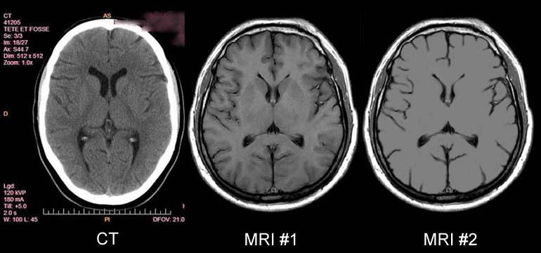
Figure 5.
Note the soft tissue detail visualized
in the MRI image #1 of the brain that is not visualized
by x-ray CT technology (e.g. the pronounced white matter-grey
matter differentiation of the MRI, the clearly defined
thalamic nuclei, and the well visualized subdural layers
not visualized by CT). MRI image #2 shows an image of
the brain where all the MR signals
of the brain tissue are the same. (i.e. no signal
difference from the TISSUES
of the BRAIN.
No grey matter -white matter differentation, no caudate
nucleus, no putamen, no thalamus.) In
the absence of the MR signal differences of the normal
tissues discovered by Damadian
(Fig.6, Fig.9) the image detail
of normal human anatomy is missing (MRI #2).
NO SIGNAL DIFFERENCES:
THE IMAGE IS A BLANK
THE SIGNAL MAKES THE IMAGE !!
NO SIGNAL DIFFERENCES: 1,
NO IMAGE !!
1. R. Damadian "Tumor Detection by Nuclear Magnetic Resonance"
Science, 19 March 1971 , Vol. 171. pp.1151 - 1152.
AND
NO ANATOMIC DETAIL
VISIBLE !!!
(Fig 5 - MRI #2)
Paul C. Lauterbur's Notebook
9-2-1971
J. Mattson and M. Simon, The Pioneers of NMR and Magnetic Resonance Imaging in Medicine: The Story of MRI
Bar-Ilan University Press, 1996, Appendix, Chapter 9, B1,B2,B3.
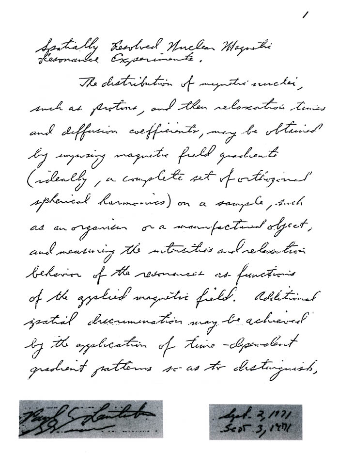
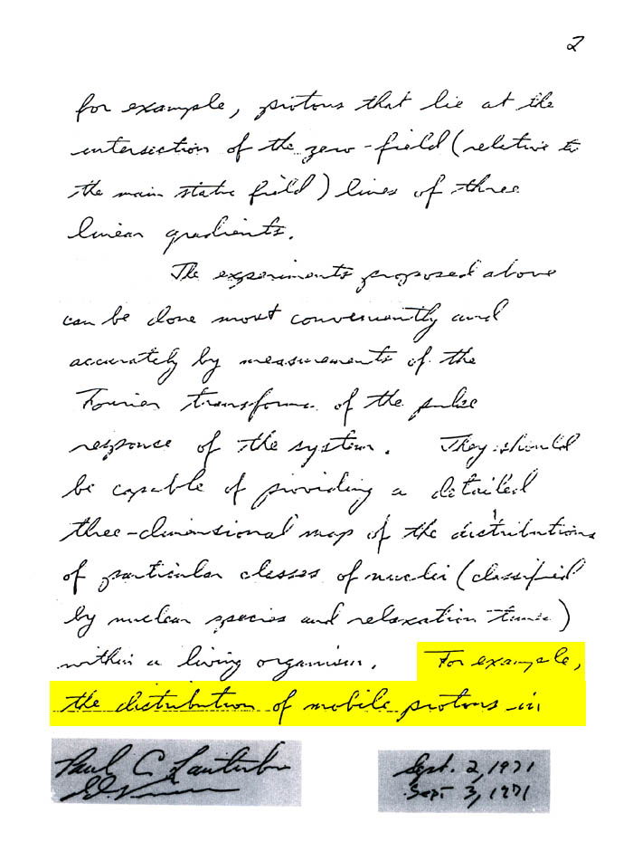
As Lauterbur published [Cancer 57, (15 May 1986), p.1899 ]
"the attention of the medical community was first attracted by the report of Damadian1 that some animal tumors have remarkably long proton NMR relaxation times.
Efforts to reproduce these results and to explore their significance were soon under way in other laboratories."
"It was measurements that I (Lauterbur) observed Saryan carring out in SEPTEMBER OF 1971 that caught my attention." [Cancer 57 (15 May 1986) p.1899.
"When Lauterbur watched Saryan successfully repeat the Damadian experiments, he viewed the procedure with great interest and was impressed by the results"2.
He (Lauterbur) Stated:
"Even normal tissues differed markedly among themselves in NMR relaxation times, and I wondered whether there might be some way to noninvasively map out such
quanties within the body " [Cancer 57, (15 May 1986), p.1899
There was nothing to MAP prior to Damadian's discovery. The NMR signal differences in normal and diseased tissues necessary to forming such a MAP were not known to exist prior to Damadian's discovery of their existence1. In their absence any such MAP would be a BLANK ! (figs. 5 and 14).
1. R. Damadian "Tumor Detection by Nuclear Magnetic Resonance"
Science, 19 March 1971, Vol. 171. pp.1151 - 1152.
2. J. Mattson and M. Simon, The Pioneers of NMR and Magnetic Resonance Imaging in Medicine: The Story of MRI
Bar-Ilan University Press, 1996, p.712, 714.
BUT THERE WAS AN
UNEXPECTED
FINDING !!
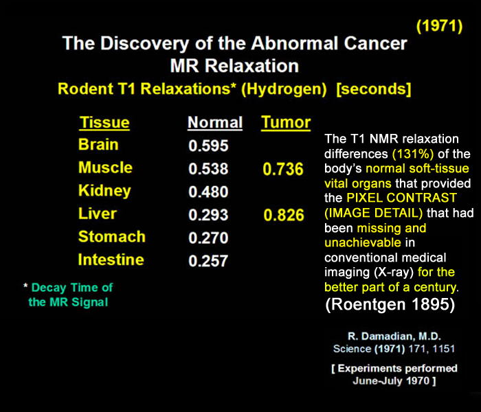
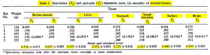
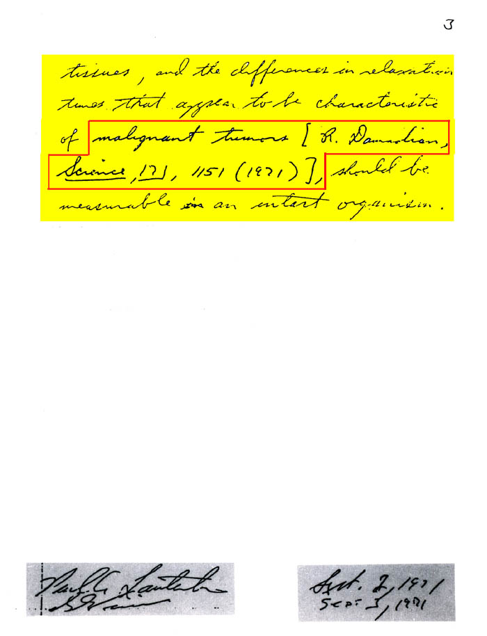
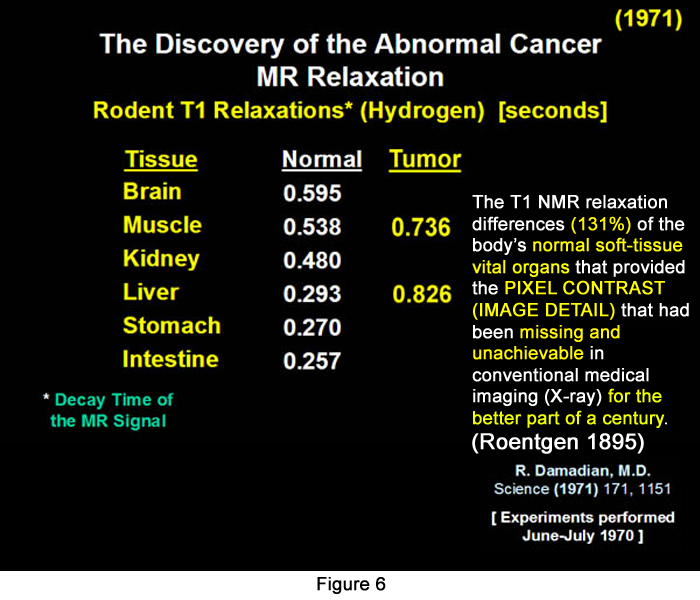
Figure 6.
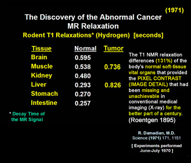
To determine if the tumor MR signal was abnormal the MR signals of the normal tissues had to be measured. Unexpectedly the normal tissues also differed markedly in their signal decay times (T1 relaxation times) e.g. the relaxation time of intestine was 257 mseconds as compared to 595 mseconds for brain (a 131% difference) with the other normal tissue relaxation times lying in between.
DR. DAMADIAN'S
DISCOVERY
OF THE TISSUE T1 DIFFERENCES OF THE BODY'S
NORMAL
VITAL ORGANS
(131%)
PROVIDED THE
PIXEL CONTRAST
(IMAGE DETAIL)
THAT HAD BEEN
MISSING
AND
RESTRICTING
MEDICAL IMAGING
(X-RAY)
FOR THE BETTER PART OF A CENTURY (ROENTGEN X-RAY 1895)
EXTRAORDINARY
IMAGE
DETAIL !!
The result of this discovery;
the pronounced relaxation time differences among the
normal tissues themselves produced an unprecented visualization
of anatomic detail in medical images that had never
been possible before by the existing x-ray imaging technology.
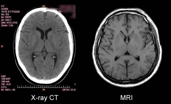
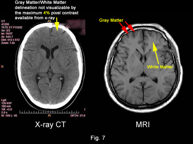
Figure 7.
As can be seen in the above T1 image of
the brain the NMR relaxation differences discovered
by Damadian made possible the imaging of the human body
at a level of detail that was unprecedented
in medical history.
The grey-white matter discrimination of the brain became visible for the first time. The thalamic nuclei, the caudate, putamen and thalamus were visualized. The Dura, layers and arachnoid layers became visible where they were not on x-ray images like CT.
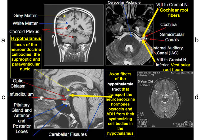
Figure 8.
Figure 8a-8d. Further examples of the
exceptional anatomic detail made visible by the DISCOVERY
of Damadian of the pronounced differences in the decay
rates (relaxations) of the NMR signals
of the body's normal tissues (Figure
6). The DISCOVERED
differences supply the pixel amplitude differences,
"
PIXEL
CONTRAST (IMAGE DETAIL)",
that produce, for the first
time in medical history, the detailed visualization
of normal human anatomy MRI is noted for. Note the visualization
of the vestibular
and cochlear nerves WITHIN
the internal auditory canal (Figure 8b) and the visualization
of the hypothalamic
tract (that transports hormones from
the brain) WITHIN
the pituitary stalk. (Figure 8c)
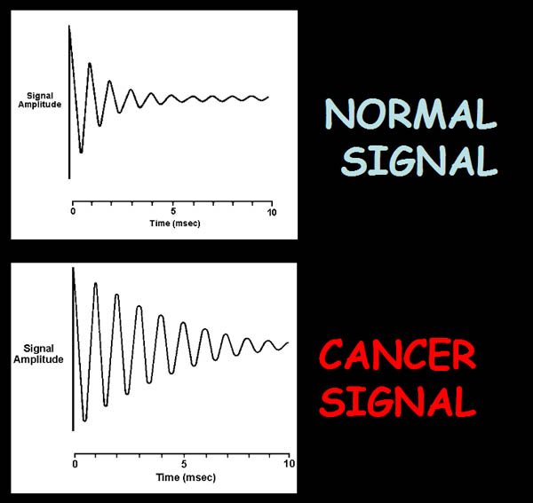
Figure 9.
Illustration of the MR signal decay rate differences of cancer and normal.
Damadian discovered
that the NMR signal
amplitudes of cancer tissue differ markedly from the
NMR signal amplitudes
of the normal tissues because of the differences in
their rate of decay.
Above is an example of the difference in the decay rate of an NMR signal from cancer tissue relative to the decay rate of a normal tissue (Tables 1 & 2). The longer the signal decay the higher the signal amplitude computed from the NMR signal. The amplitude of the tissue NMR signal sets the brightness of the pixel (picture element) in the image assigned to it as exemplified in the pixels displaying the cerebellar tumor of figure 10.
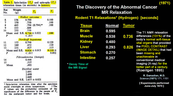
Figure 6.
The discovery
of the abnormal relaxation rates of cancers as seen
in the above malignant hepatoma (0.826)
and Walker sarcoma (0.736) T1 decay times (yellow)
Which Resulted in
Exceptional
Tumor
Definition, Visability and Detectability
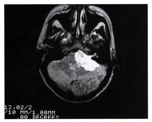
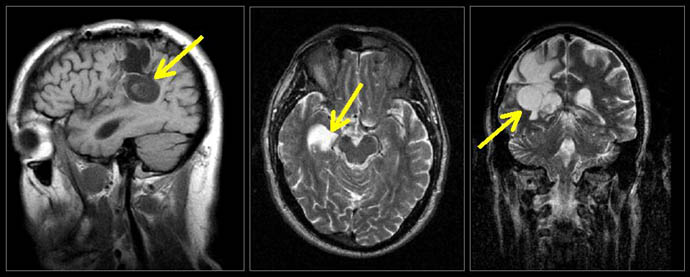
Figure 3b. An MRI Image of a
Tumor of the Brain, an Acoustic Neuroma of the Auditory
Nerve.
The striking differences in pixel brightnes, "PIXEL
CONTRAST (IMAGE DETAIL)" that separate the
tumor from surrounding normal tissue so that it can
be detected on this T2
MRI image are created
by the marked differences in T2 relaxation between tumors
and normal tissue DISCOVERED
by Damadian (tables 1, 2)
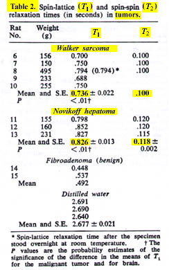
Tables 1 & 2.
R. Damadian, Tumor Detection by Nuclear
Magnetic Resonance. Science, 19 March 1971, Vol.171,
Tables 1 & 2, pp.
1151-1153. Raymond V. Damadian is the medical doctor
who first proposed scanning medical patients by NMR
(nuclear magnetic resonance, the original name of the
MRI) based on his DISCOVERY
of the principle on which all modern MRI is based —
the different NMR signals
that tissues emit in a magnetic field. The amplitude
of these signals
determines the brightness of the picture elements (pixels)
that the MRI image is composed of.
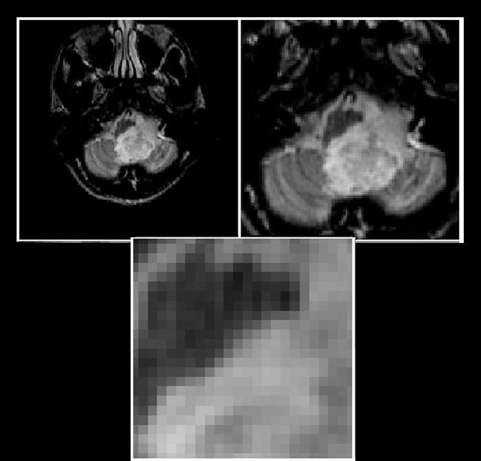
Figure 10.
A Step-wise enlargement of an MRI image of
a cerebellar tumor of the brain exhibiting the picture
elements (pixels) that
make up the image.
 |
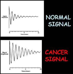 |
Figure 6. Original 1971 data in Science showing the lengthening of the decay time (relaxation time) of the NMR signal of cancer relative to normal (e.g. liver cancer 826 milliseconds (msecs) vs 293 msecs normal liver, 736 msecs Walker Sarcoma vs. 538 msecs normal muscle). The data additionally shows the pronounced differences in the NMR signal decay rates of the normal tissues (e.g. 257 msecs intestine vs. 595 msecs for brain).
|
Figure
9. He discovered
that the NMR signal
amplitudes of cancer tissue differ markedly from
the NMR signal
amplitudes of the normal tissues because of the
differences in their rate of decay.
The above is an example of the difference in the decay rate of an NMR signal from cancer tissue relative to the decay rate of a normal tissue. The longer the signal decay the higher the signal amplitude computed from the NMR signal. The amplitude of the tissue NMR signal sets the brightness of the pixel (picture element) in the image assigned to it as exemplified in the pixels displaying the cerebellar tumor of figure 10.
|
|
|
| |
T2 Image
Figure 11. Brain Tumor
|
|
  |
T2 Image
Figure 12. Tumor Metastasis to Bone
|
|
These signal
amplitude differences enabled cancer tissues (Figures
11-13) and other tissues to be visualized in MRI images
because the signal
differences generate the needed brightness differences
"PIXEL CONTRAST (IMAGE
DETAIL)" in the picture elements (pixels)
needed to visualize detail
in the MRI image.
The CONTRAST
in pixel brightness, "PIXEL
CONTRAST (IMAGE DETAIL)", allows the cancer
pixels in the image to be distinguished from the surrounding
normal pixels. (Figs 11-13)
NO SIGNAL DIFFERENCES
ALL THE IMAGE PIXELS
ARE EQUALLY BRIGHT !
NO PIXEL CONTRAST !
AND THE TUMOR IS
INVISIBLE !!
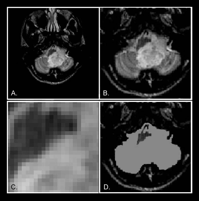
Figure 14.
The cerebellar tumor
as it would appear (14-D) with no MR signal
differences. Figure 14-D is the same image as Figure
14-B but where all MR signal
differences were eliminated and all the MR pixels therefore
had the same pixel brightness. The absence of the MR
signal differences
between cancer and normal tissue DISCOVERED
BY DAMADIAN gives the MR image
pixels equal brightness and
NO SIGNAL DIFFERENCES:
THE IMAGE IS A BLANK
THE SIGNAL MAKES THE IMAGE !!
NO SIGNAL DIFFERENCES: 1,
NO IMAGE !!
AND
THE TUMOR IS
INVISIBLE !!
(Fig 14D)
But what about
scanning for cancer?
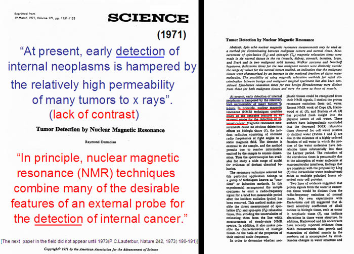
Figure 15.
the ‘832 PATENT
“Apparatus and Method
for Detecting Cancer in Tissue”
(the first ever patent on MRI)
US Patent 3,789,832
“Apparatus and Method
for Detecting Cancer in Tissue”
Filed March 1972
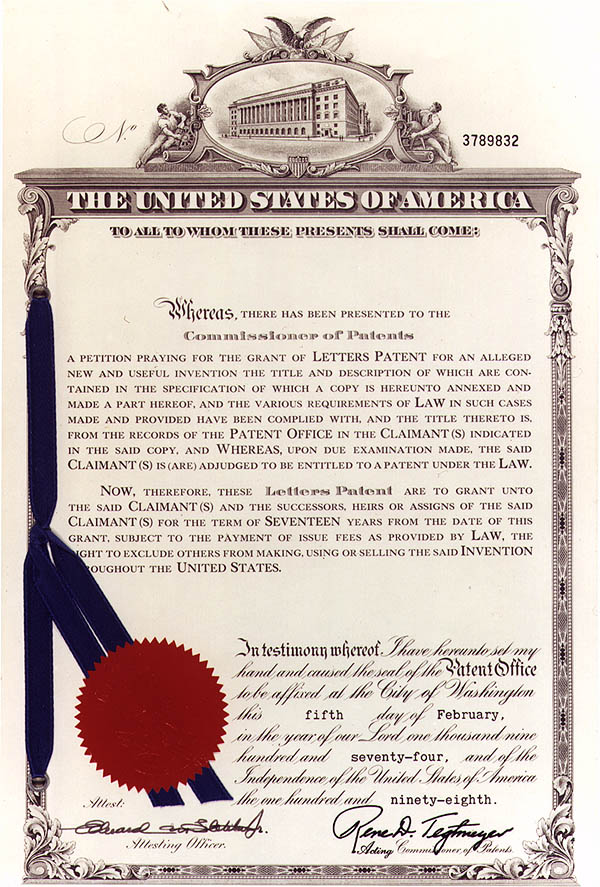
Figure 16a.
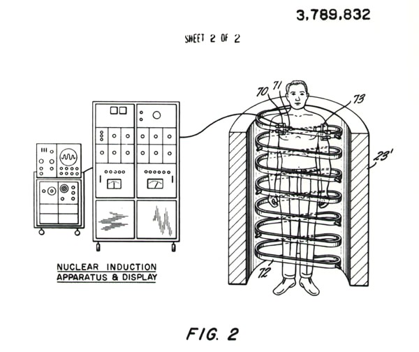
Figure 16b.
 |
(as of 2/21/13)
|
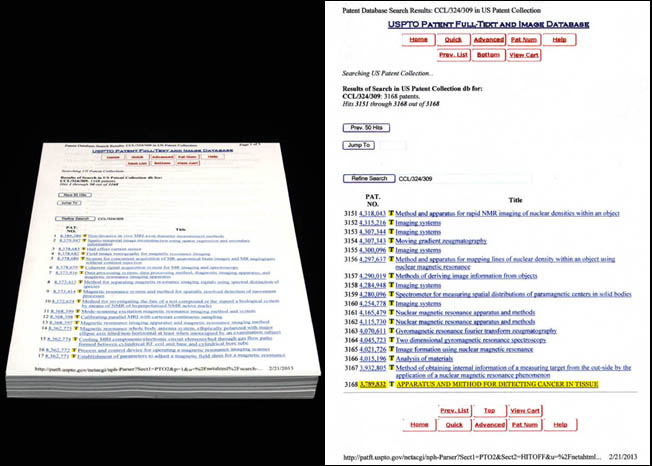
Figure 17a.
'832-The first of 4552
Patents on MRI
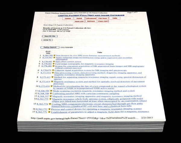
Fig. 17b.
The above is the listing of all
of the titles of the 4552 patents
(as of 2/21/13) issued for MRI
by the United States Patent Office following
Dr. Damadian's original '832 patent for MRI (filed March
17, 1972).
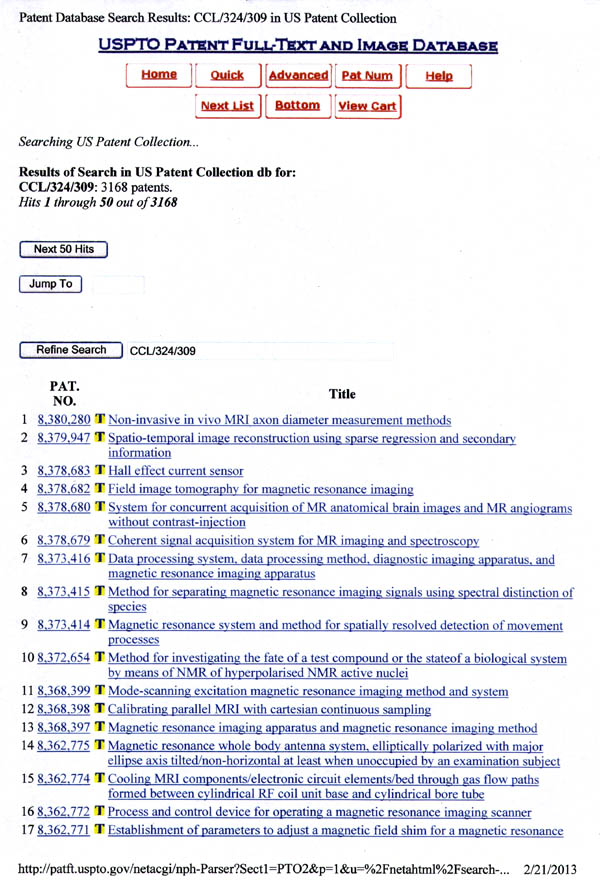
Fig. 17c.
The above is the first page of
the title list of the 4552 patents
issued for MRI by the United States Patent Office
beginning with the most recently issued patent (as of
2/21/13).
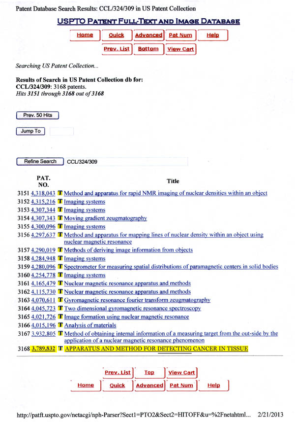
Fig. 17d.
The conclusion of
the title listing of the 4552
patents issued for MRI by the United States Patent
Office (as of 2/21/13) following
Dr. Damadian's original patent for MRI that inaugurated
the MRI industry. [U.S. Patent
"Apparatus and Method For Detecting
Cancer in Tissue". #3,789,832. Filed March 17,
1972].
US Patent 3,789,832
Upheld by the United
States Supreme Court
Oct 6, 1997
But not always smooth sailing !
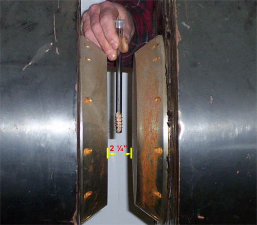
Figure
18.
The standard 23 year old 2 ¼"
NMR Test-Tube Analyser used by chemists
for ascertaining the molecule composition of aqueous
solutions prior to Dr. Damadian's discovery.
The standard NMR(MR)
test-tube analyzer, utilized by NMR spectroscopists
at the time had a two and 1/4
inch gap between the magnet poles to accept test-tube
samples. It was the only NMR apparatus in existence
at the time Dr. Damadian did his original NMR (MR) test-tube
analyses of normal and cancerous tissue samples to see
if a disease (cancer) differentiating NMR signal
could be experimentally demonstrated that would enable
his concept of a cancer detecting NMR(MR) body scanner
to proceed.
Telling
someone looking at this apparatus that it should be
used to scan the human body was regarded as absurd.
The giant magnets to do it did not exist. The rf antennas
needed to accomplish detecting a less than 1mm tumor
inside the body also did not exist. They were a major
concern.
The sample tube
is non-invasively wrapped with an external transmitter-receiver
coil to stimulate and receive nuclear resonance signals
from the sample.
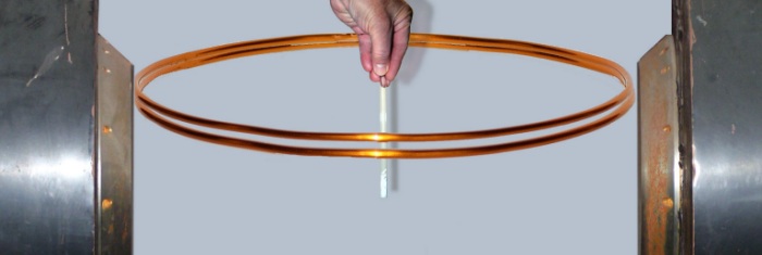
Figure 19a.
With the same tissue
sample as in the above illustration but now 10"
removed from the proposed MR antenna envisioned for
a body MR scanner, and where the MR signal
itself was not all that strong and readily lost by the
slightest mispositioning of the sample within the magnet
the prospect of successfully acquiring an MR signal
with an external antenna from a 1mm tissue sample deep
within the human body was a
major uncertainty.
At
the time the idea
(1971) of taking a 2¼ inch test-tube analyzer
and turning it into a scanner of the live human body
was deemed absurd.
“THEREFORE ANY FURTHER DISCUSSION
ABOUT SCANNING THE HUMAN BODY BY
NMR IS VISIONARY NONSENSE ” |
|
This was the conclusion of an NMR scientist of the John
Hopkins Medical Center, one of the three NMR scientists (Raymond Damadian, Carlton Hazlewood and Donald Hollis)
granted a research contract by the National Cancer
Institute's (NCI) Cancer Diagnosis Project in
1976, after his successful repeat of Dr. Damadian's
demonstration of the prolonged relaxations of
the NMR signals of cancerous tissues and his additional
observations that non-malignant diseased
tissues also had prolonged NMR relaxations.
He had, however, overlooked that both cancerous
and non-cancerous diseased
tissue NMR signals were markedly
prolonged relative to normal, making the PIXELS
of BOTH diseased
tissue types conspicuously brighter
than normal on a medical image for the first time
and eminetly visible by MRI.
Professors of John Hopkins
Medical Center present at the conference immediately
disagreed with their colleague's "visionary
nonsense" claim stating that "Now Doctor,
just tell us where to put the needle we are way
ahead of where we are today". They further
refuted his declaration that it was "visionary
nonsense".
|
Figure 19b.
|
| At a subsequent
and unrelated litigation the infringer of Dr. Damadian's
patent made the same argument, that the elevated
NMR relaxation times for cancer were also elevated
in other diseased tissues
that were non-cancerous. FONAR's attorneys responded,
" Ladies
and gentleman of the jury are you going to punish
the guy because his original discovery
detects MORE
DISEASE than
he originally envisioned ? "
Dr. Damadian and Fonar prevailed. |
| |
At
a subsequent conference of NMR scientists where
Dr. Damadian had been invited to present his NMR
findings in cancer, the Moderator of the NMR conference,
at the conclusion of Dr. Damadian's presentation
stood to ask |
“ NOW DOCTOR HOW FAST DO YOU
PROPOSE
TO SPIN THE PATIENT ?”*
|
* (Spinning the test tube sample at high rpm was a standard in NMR spectroscopy for overcoming the magnetic field inhomogeneities that the protons of the test tube sample were exposed to) |
|
Using
Brookhaven National Laboratories magnet design software,
MAGMAP, that had been provided by the scientists from
Brookhaven National Laboratories, Dr. Gordon Danby,
Dr. Hank Hsieh and John Jackson, who had joined Dr.
Damadian as Fonar consultant employees to try to build
the first NMR (MRI) scanner of the live human body
that Dr. Damadian was trying to construct, Dr.
Damadian calculated that 30 miles (150,000 feet)
of SUPERCONDUCTING Niobium
Titanium (NbTi) magnet wire was needed to produce
the
5,000 gauss magnetic
field he had estimated was necessary to achieve a
successful NMR scan of a live human body. "At
the existing wire price of $1
per foot, the projected wire cost was $150,000
and I only had $15,000
in my budget"
Additionally
the Niobium Titanium (NbTi)
superconducting wire needed the making of SUPERCONDUCTING
joints between successive lengths of wire in order
not to undo the SUPERCONDUCTIVITY
of the final magnet.
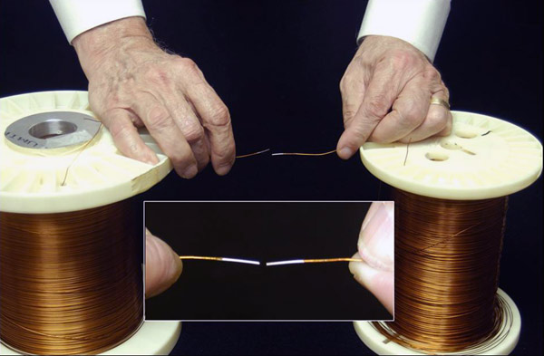
The
NbTi liquid helium superconducting
wire was not available from suppliers on a single
spool. It required SUPERCONDUCTING
joints to be made between successive lengths of the
currently available NbTi
wire.
I called
Steve Lane, my sales representative at Westinghouse,
the source of the SUPERCONDUCTING
NbTi wire I was considering, and asked him
if he would teach me how to make the
superconducting joints I needed. His response
was " What are you doing Dr. Damadian? Are you
going into competition with Westinghouse? "
I said " no no Steve I'm trying to make
a superconducting NMR
magnet that would be big enough
to achieve an NMR scan of the live human body "
Steve
responded by saying "it's good that you levelled
with me Dr. Damadian. I can share with you something
that no one else knows as yet. Westinghouse is going
out of the business of making superconducting
wire. I have about 30
miles of superconducting wire I can let you
have for 10¢ on the dollar"
I was
dumbfounded. With no prior knowledge from me, at Westinghouse
or anywhere else, that I wanted
to build an NMR magnet big enough to scan the human
body, Westinghouse after
23 years of manufacturing their Niobium
Titanium wire was SUDDENLY
discontinuing their 23 year
old manufacturing of NbTi
wire, and had in their warehouse EXACTLY
the amount of wire I had CALCULATED I NEEDED (30
miles) and would let me have it for THE
EXACT AMOUNT I HAD IN MY BUDGET.
I was
amazed by this extraordinary coincidence of Westinghouse
ceasing to make the wire they had been making for
23 years at the precise
instant I needed it, and at
the exact length I needed (30
miles of wire) and at a price of " 10¢
on the dollar " that matched the exact
amount I had in my budget ($15,000).
I happened
to mention this exceptional
coincidence of the wire's sudden availability
from Westinghouse at 10¢
on the dollar at the exact instant I needed
it, to my wife's mother and father, Amy and "Bo"
Terry (both evangelical christians).
My wife's
mother responded " That's
no coincidence Raymond " Ever since your
Dad and I learned of your desire to build your scanning
machine we've been praying for you " " This
is not a coincidence. It's an answer to prayer !
"
From
which I concluded that this sudden availability of
the magnet wire AT 10¢
ON THE DOLLAR AT THE
VIRTUAL INSTANT I NEEDED IT, was not an accident
but that JESUS
from my mother's prayers HAD
JUST GIVEN
MANKIND THE
MRI
!
" I WISDOM DWELL WITH PRUDENCE AND FIND OUT KNOWLEDGE OF
WITTY INVENTIONS "
(Proverbs. 8:12 - KJV)
" FOR THE LORD GIVETH WISDOM:
OUT OF HIS MOUTH COMETH KNOWLEDGE AND UNDERSTANDING "
(Proverbs. 2:6–8 - KJV)
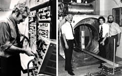
Construction of the First Human MR Scanner, Indomitable, Begins.
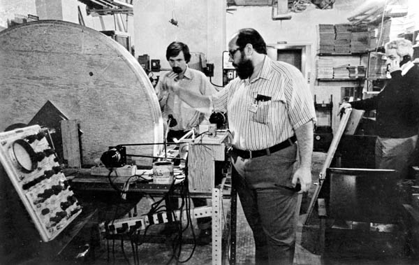
Figure 20a.
Michael Goldsmith
and Michael Stanford winding one of the two Niobium
Titanium (NbTi) superconducting magnet coils built for
Indomitable.
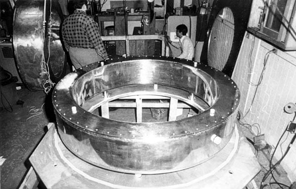
Figure 20b.
Michael Goldsmith
and Nean Hu with the liquid helium cryogen chamber that
housed the NbTi superconductiong magnet coil.
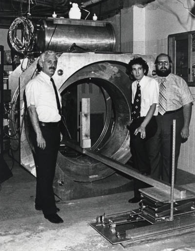
Figure 20c.
Left to Right, Raymond Damadian,
Larry Minkoff and Michael Goldsmith alongside "live
magnet" Indomitable
with the iced liquid helium port on the top of the magnet
alongside of the two helium cooling liquid nitrogen
ports.
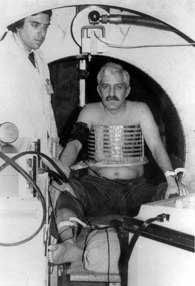
Figure 20d.
Dr. Damadian in
Indomitable for the first
attempt at a human MR scan with his chest surrounded
by the largest diameter antenna (14" diameter)
that Dr. Goldsmith had been able to build at the time
that could still successfully generate any MR signal
from an interior sample. Also pictured is an adjacent
cardiac defibrillator to counter any emergencies that
might arise and a cardiologist to administer it if necessary.
The scan attempt on Dr. Damadian failed. All that was
obtained was a normal EKG.
"VISIONARY NONSENSE"
had just turned into a
SAD REALITY !
The Goldsmith hypothesis
for the failed scan was Dr. Damadian was "too
fat" for his coil and was loading
the coil's impedance.
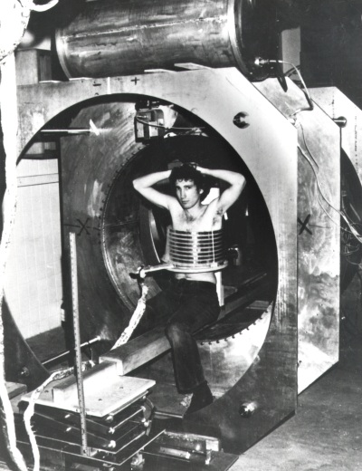
Figure 20e.
Larry Minkoff
finally gets into Indomitable
(weeks later) to test the Goldsmith "too
fat" hypothesis.
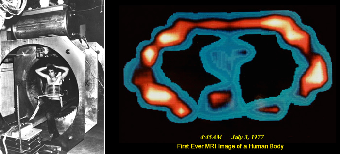
4:45 AM
July 3, 1977
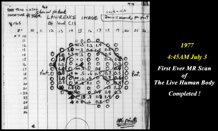
Figure 21.
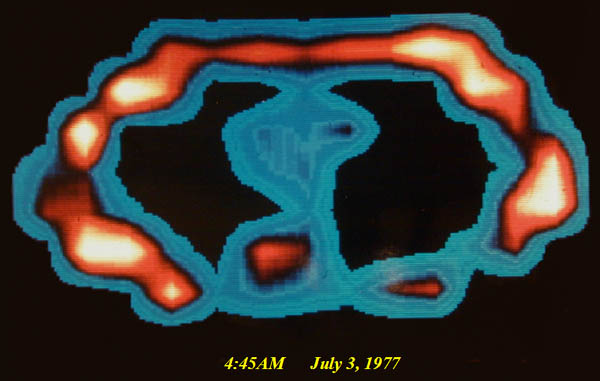
Figure 22.
FIRST
EVER MRI IMAGE OF THE LIVE HUMAN BODY !!
A cross-section of L. Minkoff's chest at the level of
T-8 showing chest walls, lungs, heart, aorta and vertebra,
and the suggestion of cardiac chambers within the heart
that was initially put down as too good to be true.
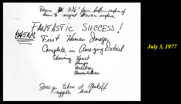
Figure 23.
"VISIONARY NONSENSE"
HAD JUST BEEN TURNED
into
REALITY !
The exhilaration
of team Indomitable at
4:45 AM July 3, 1977.
Hallelujah !
The
WILD IDEA
( "Visionary Nonsense" )
( turning a 10 mm test tube analyzer, Fig. 18,
into a scanner of the LIVE human body )
HAD
MIRACULOUSLY
just become
THE NEW REALITY ! ! ! ! !
A 2¼ inch test-tube analyzer (Fig. 18)
of
23 years duration
had just been
TRANSFORMED !
into a scanner of the live human body
MRI WAS BORN !!!
2nd MAJOR DISCOVERY
Exceptional Anatomic Detail in Medical Images for the First Time in History
Clear Visualization of the Body's Vital Organs
T1 and T2
Revolutionize Medical Imaging
The T1 and T2* tissue differences
DISCOVERED by Damadian
Revolutionize Medical Imaging
ANATOMIC
DETAIL
visualized in the
body's critical vital organs
for the first time in
medical history
*
Damadian's discovery
of their profound differences within the vital life-giving
tissues of the human body that enabled his idea
of the NMR scanning of the live human being. (Damadian,
R. "Tumor Detection by Nuclear Magnetic Resonance",
Science, March 19, 1971, 171, 1151-1153)
PIXEL CONTRAST
(difference in pixel brightness of adjacent image pixels)
A typical medical image is constructed from PICTURE ELEMENTS
designated PIXELS (Fig 14C) in medical imaging nomenclature.
Fig. 14
A typical 256 X 256 medical image therefore is a composite of 65,536 picture
elements
(pixels-Fig 14c)
Accordingly
the power to visualize
DETAIL
in the body's
CRITICAL SOFT TISSUE VITAL ORGANS
(e.g. Brain, Heart, Muscles, Kidney, Liver, Spleen, Pancreas, Intestines)
therefore RESTS ENTIRELY on the power of the imaging technology to generate the
PIXEL CONTRAST
needed to visualize
IMAGE DETAIL
in the body's vital tissues. The existing x-ray technology for visualizing
IMAGE DETAIL
in the body's
CRITICAL VITAL ORGANS
had been severely lacking in its power to generate the
PIXEL CONTRAST
needed to visualize
IMAGE DETAIL
in the body's
CRITICAL
VITAL SOFT-TISSUE ORGANS
Dr. Damadian's discovery of the NMR signal
differences of the body's vital tissues, the signal amplitude differences generated by their tissue NMR signal decay time differences
(their NMR "relaxation time" differences) provided a
131% PIXEL CONTRAST
(Fig 6, Tables 1 & 2) for visualizing
IMAGE DETAIL
in the body's VITAL SOFT-TISSUE ORGANS as compared to a maximum
4% PIXEL CONTRAST
available from X-ray. This provided the
PIXEL CONTRAST
needed to visualize
IMAGE DETAIL
in the body's
CRITICAL
SOFT-TISSUE VITAL ORGANS
that had been missing from traditional medical imaging (x-ray imaging)
for the better part of a century
(Roentgen 1895)
Fig.6
(Tables 1 & 2)
SATISFACTORY VISUALIZATION,
for the
FIRST TIME
IN MEDICAL HISTORY,
of the
CRITICAL SOFT TISSUE DETAIL
of the
LIFE GIVING
VITAL ORGANS
OF THE HUMAN BODY !!!
(Brain, Heart, Spine, Muscles, Liver, Kidney, Spleen, Pancreas, Intestines ...)
EXCLUDED BY THE NP !
Damadian's discovery
of the "signal that makes the
image" provided another
VITAL
DISCOVERY.
It provided,
FOR THE FIRST TIME IN MEDICAL HISTORY,
the power to
achieve clear visualization of the body's
VITAL ORGANS.
Prior to the
advent of MRI, medical imaging from its x-ray inception
in 1895 was uniquely deficient in its ability to achieve
satisfactory (and necessary)
visualization of the life-sustaining organs of the
human body (Brain, Heart, Muscles,
Kidney, Liver, Spleen, Pancreas, Intestines ...).
X-ray technology was significantly limited in its
ability to produce the
PIXEL CONTRAST
needed to visualize
IMAGE DETAIL
in the body's soft-tissue vital organs due to the
small x-ray transmission differences of
x-ray radiation across the body's soft tissues (unlike
the transmission difference between bone and soft
tissue that generate pronounced image contrast on
x-ray images and excellent visualization of bone on
x-ray images). The difference in x-ray transmission
across the body's soft tissues, and therefore the
ability to generate the necessary PIXEL
CONTRAST in x-ray images to visualize
IMAGE DETAIL in the body's
VITAL
SOFT TISSUE ORGANS,
was severely
limited to a maximum PIXEL CONTRAST
of 4% by x-ray
imaging technology.
The
picture element
(pixel) is the smallest
display element in a medical image. A typical 256
x 256 pixel medical image
is composed of 65,536 pixels
(256 x 256). The ability to visualize detail in an
image is dependent on the capability of adjacent picture
elements (pixels)
in an image to display differences in brightness (PIXEL
CONTRAST)
that reflect the differences in anatomy of the adjacent
anatomic structures.
Consequently the ability of
the pixels of an image
to exhibit differences in brightness (PIXEL
CONTRAST)
and visualize ANATOMIC
DETAIL in the image is dependent on the power
of the imaging technology to generate differences
in the brightness in the picture
elements (pixels)
that make up the image.
Accordingly
the magnitude of the PIXEL CONTRAST
achievable by the pixels
of an MRI image reflects the power of the image to
visualize ANATOMIC DETAIL. Consequently the marked
differences in the T1 and T2 tissue NMR relaxations
(a
131% PIXEL
CONTRAST: Fig 6, Tables 1 &2)
achieved
by MRI, as compared to the maximum of a
4% PIXEL CONTRAST
achieved by X-ray, accomplished
DETAILED VISUALIZATION
of
the body's
CRITICAL
LIFE-GIVING VITAL ORGANS
(Brain, Heart, Muscles, Kidney, Liver, Spleen, Pancreas,
Intestines ...)
FOR THE FIRST TIME
IN MEDICAL HISTORY
THE EXCEPTIONAL ANATOMIC DETAIL
ENABLED
by
THE TISSUE NMR RELAXATION
DISCOVERIES
of
Dr.
Damadian
that was UNPRECEDENTED
IN MEDICAL HISTORY and
had been beyond reach in medical imaging for nearly
a century (Roentgen 1895).
UNPRECEDENTED
IMAGE DETAIL
WITH NO
IONIZING RADIATION !
Figure 8.
T1 and T2 Medical
Images of the Brain
Figure 8
Figure 8a-8d. Further examples of the exceptional anatomic detail made visible by the DISCOVERY
of Damadian of the pronounced differences in the decay rates (relaxations) of the NMR signals
of the body's normal tissues (Figure 6). The DISCOVERED
differences supply the pixel amplitude differences,
"PIXEL CONTRAST (IMAGE DETAIL)",
that produce, for the first time in medical history, the detailed visualization of normal human
anatomy MRI is noted for. Note the visualization of the vestibular and cochlear
nerves WITHIN the internal auditory
canal (Figure 8b) and the visualization of the hypothalamic tract (that transports
hormones from the brain) WITHIN the pituitary
stalk. (Figure 8c)
Damadian's
discovery
of the NMR signal differences that provided the MRI
visualization and detection of diseased tissue (Fig.
6, Tables 1 & 2) overcame this deficiency also. He
discovered
that the NMR signal
differences (T1
and T2 signal relaxation differences) among
the body's NORMAL soft
tissues (Brain, Muscle, Kidney, Liver, Stomach,
Intestine) were also pronounced
(Fig. 6, Tables 1 & 2) (e.g. small intestine 257msecs,
brain 595 msecs, a 131%
difference, Fig. 6). These large
NMR signal differences in the body's NORMAL
soft tissues, discovered
by Damadian, overcame, for the first time,
medical imaging's longstanding deficiency in its visualization
of the body's
VITAL
soft tissue normal organs (Figs. 7, 8).
The newly discovered
marked differences in
the proton NMR relaxation
times of the body's VITAL
soft tissues (131%)
enabled medicine to SURMOUNT
its historic deficiency, it's inability to generate
the necessary image
CONTRAST (PIXEL
CONTRAST)
crucial to visualizing the needed ANATOMIC
DETAIL of the body's
CRITICAL LIFE-GIVING VITAL ORGANS.
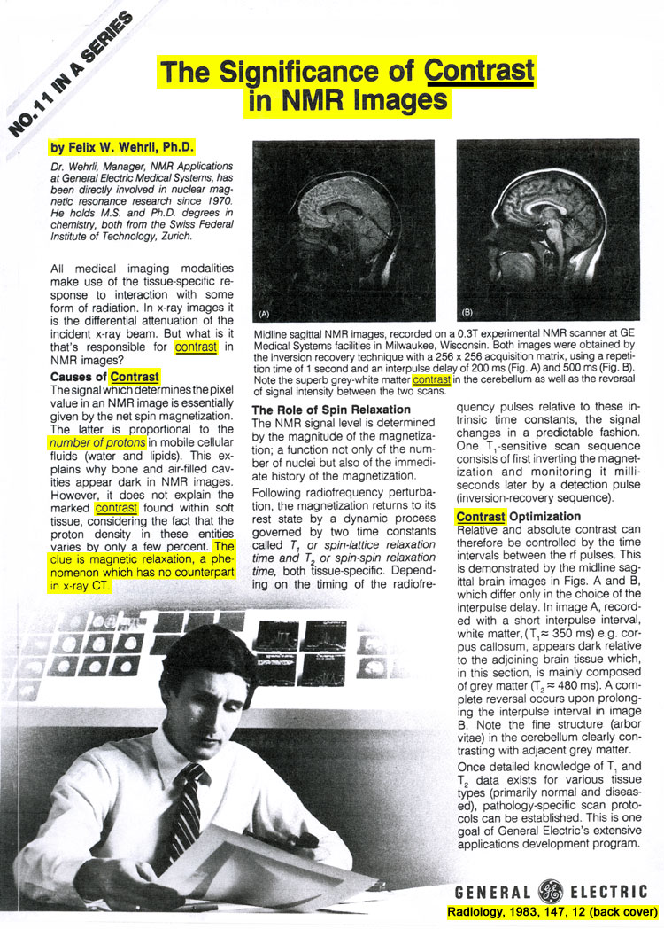
As Dr.
Felix Wehrli PhD, MRI Imaging scientist and
manager of the NMR Applications Division of the General
Electric Company in his 1992 publication
(Wehrli, F.W. Physics Today, June 1992, 34-42)
REPORTED
"
The Origins and Future of Nuclear Magnetic Resonance
Imaging "
(regarding
the fundamental importance of the discovery
by Dr. Damadian
of the abnormal NMR relaxation times of diseased tissue),
"
it was recognized early on
that
in most diseased tissues, such as tumors,
the relaxation times are
prolonged
(R.
Damadian, 1971, Science 171, 1151)."
"This
difference provides the basis for image CONTRAST
between normal and pathological tissues ".
(Wehrli, F.W. Physics Today, June 1992, p38)
Wehrli had previously reported
in
"
THE SIGNIFICANCE OF CONTRAST IN NMR IMAGES ",
that
"
the number of protons... does
not explain the marked CONTRAST
found within soft tissue, considering the fact that
the proton density in these entities varies by only
a few percent.
THE CLUE IS
magnetic relaxation, a
phenomenon which has no counterpart in x-ray CT
"
(Wehrli, F.W. Radiology
1983, 147, 12 (back cover).
T1 and T2 Imaging
Without Dr. Damadian there is no T1 or T2 in the practice of medicine.
90%
of all MRI images acquired today
are either
T1 weighted or T2 weighted MRI images
(e.g. T1 axial lumbar MRI, T2 coronal brain MRI)
T1 and T2 would not exist in the practice of medicine
except for Dr. Damadian's DISCOVERY of the
CHANGES
in the
T1 and T2 NMR SIGNALS OF THE BODY'S TISSUES that are created by the pathologic tissue changes generated by disease.
T1 and T2
existed
NOWHERE
in the
PRACTICE OF MEDICINE
until
AFTER
Dr. Damadian's
DISCOVERY
of the
ALTERATION of T1 and T2
by
DISEASE
and their
POWER
for
DETECTING DISEASE
in the
BODY'S LIVE TISSUES ! :
i.e. their POWER to generate the necessary
PIXEL CONTRAST
(IMAGE DETAIL)
in images of the body's soft-tissue vital organs that had been missing
and unachievable in conventional medical imaging
for the better part of a century
(x-ray).
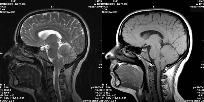 T2
sagittal MRI image of the brain T1
sagittal MRI image of the brain
T2
sagittal MRI image of the brain T1
sagittal MRI image of the brain
Dr. Damadian's
DISCOVERY
of the
ABNORMALITIES
of the
T1 and T2
TISSUE NMR SIGNALS
GENERATED
by
TISSUE DISEASE !
In NMR, two ways exist with which to characterize
the nuclear signal response of the resonating
atomic nucleus. You can analyze either the frequency
dependence of the nuclear signal (NMR spectroscopy)
or its time dependence, i.e., its rate of decay
(relaxation time). Both characterizations are
manifestly informative but until the Damadian
discovery
of the key sensitivity of the decay time of
the nuclear resonance signal to the existence
of disease within medical tissues, the overwhelming
majority of the uses of NMR technology (prior
to its medical application by Damadian) were
its chemical applications (NMR spectroscopy)
for determining, non-destructively, the molecular
composition of organic molecules.
Thus the nuclear resonance signal has two properties, its frequency response to excitation and the time dependence of the nuclear signal's response to excitation, that are very informative regarding the chemical composition of molecules and their surroundings.
Regarding the time dependencies of the nuclear resonance signal, there are two, one of which is eminently visible in the oscilloscopic display of the signal as a function of time (T2) (Figs. 4a and 4b), and a second, usually not visible, that is a measure of the time required by the stimulated nucleus to return to its equilibrium state after its excitation (T1). The times of these two time-dependent nuclear signal responses, T1 and T2, commonly differ markedly, e.g., T1 = 538msecs in muscle, T2 = 55msecs in muscle (Table 1).
The Time Dependent Decay (relaxation) of the Nuclear Resonance Signal
In the time dependence characterization of the nuclear resonance signal, two time dependent phenomena are experimentally encountered, the rate of dissipation (T1) of the stimulating energy (the r.f. pulse) after it has been applied and generated the NMR signal and the rate at which the individual signals generated by each resonating nucleus within the sample dephase and destructively cancel the composite resonant signal of the individual nuclear resonance signals (T2).
The T1 Relaxation
The T1
decay rate (its T1 relaxation time) is the time
of dissipation, (e.g., muscle T1 = 538msecs.
Table 1), to the surroundings (the "lattice"),
of the r.f. pulse energy used to stimulate the
NMR signal; i.e., the "spin-lattice relaxation
time": the "thermal relaxation time".
The T2 Relaxation
The T2 decay rate (its T2 relaxation time) is the time rate of decay of the live NMR signal (Figs. 4a and 4b) actually observed on the oscilloscope, e.g. muscle T2 = 55msecs. Table 1. It is the time for the destructive interferences of the phase incoherencies of all of the NMR signals generated by the individual atoms within the sample to reduce the magnitude of the observed composite signal to zero (Figs. 4a and 4b).
Noteworthy
is the fact that the great majority of all MRI
images acquired today are either T1 or T2 weighted
images (90%). The use of such T1 and T2 relaxation
dependent images explicity exploit the benefit
of the Damadian discovery
2 that the T1 and T2 signal relaxations
exhibit the most pronounced (and therefore most
visible) discriminations among the body's normal
and diseased tissues as compared to their differences
in hydrogen content (proton density images).
80 - 90% of all patient MRI scans performed
today utilize either T1 (T1 weighted) or T2
(T2 weighted) imaging protocols. With approximately
15 million patient MRI scans being performed
each year in the U.S. and an equal number being
performed each year in the rest of the world,
30
million T1 (or T2) patient MRI scans are being
performed worldwide each year
using the original
T1 and T2 tissue MR (NMR) signal
differences discovered
by Damadian.
These MR
signal
T1 and T2 differences of diseased tissue (EXCLUDED
by the NP3)
as well as the significant
T1 and T2 differences of the MR signals
of normal tissues (EXCLUDED
by the NP), are THE
SIGNALS THE MRI MAKES THE IMAGE WITH
(the signals
used by the MR scanner to construct the MRI
images). Without these tissue MR signals
differences discovered
by Damadian that are used to construct
the MRI image, there would be no MRI today.
With each
T1 or T2 scan consisting of approximately 15
image slices (15 images/scan) per patient scan,
30 T1 or T2 image slices are being acquired
world-wide
each year from each patient: i.e. the better
part (90%) of 1
BILLION
T1 and T2 MRI images
(900
MILLION images) are being acquired
each year throughout the world, for the benefit
of humanity, thanks to the tissue T1 / T2 signal
relaxation differences in the body's vital organs
discovered
by Damadian.
Indeed,
the MR signal
T1 and T2 differences of the MR signals
of diseased and normal tissues discovered
by Damadian (and
EXCLUDED by the NP) make all the
T1 (T1 weighted) and the T2 (T2 weighted) images
of MRI.
While the
imaging techniques awarded the NP have long
been replaced4,
the T1 and T2 images based on Damadian's discovery
(and
EXCLUDED by the NP) continue to
produce every MRI patient examination in the
world that is acquired today. Indeed, it is
difficult to imagine, for example, how the T2
MRI scan originated by the Damadian discovery
(and
EXCLUDED by the NP) that visualizes
all diseased tissues wherever they might occur
within the human body can ever be replaced.
It is in fact hard to envision how it will not
persist as the forever
component of the patient MRI Examination while
the initial techniques5
to make use of the tissue MR (NMR) signals
discovered
by Damadian to make the image have long been
replaced6.
|
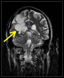
Figure 11.
T2 MRI visualization of a tumor of the brain made possible
by the discovery
of Damadian of the abnormal T2 (and T1) MR (NMR) relaxations
of cancerous tissue.
| |
|
| 2 |
Damadian, R., Tumor Detection by Nuclear Magnetic Resonance. Science, 171:1151-1153, 1971 |
| 3 |
|
| 4 |
By phase contrast frequency scanning |
| 5 |
Lauterbur, P.C. (1973) Nature, 242:190-191 (NP 2003) ( PhD. 1962): Garroway, A.N., Grannell, P.K., Mansfield P. (1974) ( PhD. 1962) J. Phys. C: Solid State Phys., 7:L457-L462, (NP 2003) |
| 6 |
Regarding
Paul Lauterbur and Peter Mansfield, recipients
of the NP, both had been
working in the field of NMR for nine (9) years
prior to Damadian's discovery5.
Neither conceived of the idea
of scanning the human body by NMR nor provided
the means to bring it about by discovering the
tissue NMR signal
differences that made it happen. Neither
did anything in the field of NMR scanning for
nine (9) years until AFTER
Damadian first conceived of the NMR body scanner
(1969) and published the means (the signals)
to accomplish it (1971). |
2nd MAJOR DISCOVERY
QUESTION: What did MRI bring to medical imaging that had
been lacking
for the better part of a century
(W. Roentgen X-ray 1895) ?
ANSWER:The power to visualize
Unprecedented
Medical Image Detail in a medical image for the first time in medical history.
(Fig 7 & 8)
Figure 7
Figure 8
QUESTION: What did Dr. Damadian's DISCOVERY
provide to medical images that SURMOUNTED the inability of existing medical imaging technology
(x-ray) to visualize DETAIL in medical images?
ANSWER: PIXEL CONTRAST
QUESTION: What delivered the
PIXEL CONTRAST ?
ANSWER: T1 and T2.
Picture Elements (PIXELS): the structural component of the medical image
A typical MRI image is composed of 65,536 PICTURE ELEMENTS (PIXELS), i.e. a rectangular 256 X 256
PIXEL MATRIX (Fig 10) consists of 256 pixel rows and 256 pixel columns.
Fig 10.
The power to visualize detail in any pixel image resides in the power of the individual image pixel to generate differences
in pixel brightness (PIXEL CONTRAST)
(Fig 14a, b, c, d)
(Fig 14a, b, c, d)
The differences in the tissue NMR relaxation times (T1 and T2) of the body's healthy tissues (131%),
DISCOVERED by Dr. Damadian (Tables 1 & 2, Fig 6), provided
the signal amplitude differences that generate the pronounced brightness DIFFERENCES of the MRI
image pixels and produce a 131% PIXEL CONTRAST for the visualization of ANATOMIC
DETAIL in MRI medical images that had been limited to a maximum PIXEL CONTRAST
of 4% for visualizing anatomic detail by x-ray.
Note the marked advance in
IMAGE DETAIL
achieved by the MRI.
Note how the 131% PIXEL CONTRAST achieved by the MRI as compared to the maximum
4% PIXEL CONTRAST provided by x-ray visualizes and distinguishes the GRAY MATTER OF THE
BRAIN FROM THE WHITE MATTER OF THE BRAIN (Fig 7) which cannot be achieved by the maximal 4%
PIXEL CONTRAST available from x-ray.
( EXCLUDED BY THE N.P.)
BEST IMAGE QUALITY IN MEDICAL HISTORY !
The SIGNAL MAKES THE IMAGE !
The NMR signal differences of the body's vital organs
DISCOVERED by Dr. Damadian
(Fig 6, Tables 1 & 2)
SURMOUNTED a Historical Major Deficiency in Medical Imaging ,
Unsatisfactory Visualization of the Body's Critical Vital Organs (Brain, Heart, Kidney, Liver, Intestine
...
Because of Damadian's profound discovery,
the
PRONOUNCED NMR signal differences
of the
body's CRITICAL TISSUES
(Fig 6, Tables 1 & 2),
its
life-sustaining soft tissue vital organs (brain, heart, liver, spinal cord, intestines, etc)
can be
visualized in detail in a medical image.
for the
FIRST TIME IN MEDICAL HISTORY !!!
The NMR signal differences of the body's vital organs,
DISCOVERED by Dr. Damadian
(Fig 6, Tables 1 & 2), supply the missing anatomic detail that had been lacking in medical images
for the better part of a century
(Roentgen 1895)
A 131% difference in soft tissue pixel contrast (brain vs. small intestine,
Fig 6) replaced a maximum 4% difference in soft tissue pixel contrast available from x-ray
 |
 |
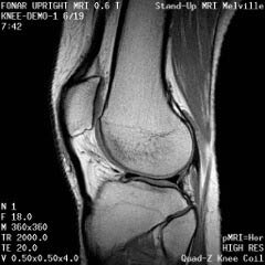
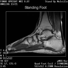
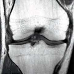
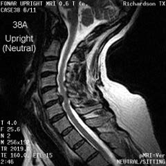
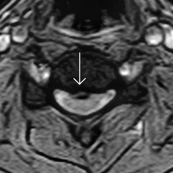
Cervical Spine |
Upright Neutral |
Upright Extension |
Unsuspected Disc Herniation in Extension |
| |
 |
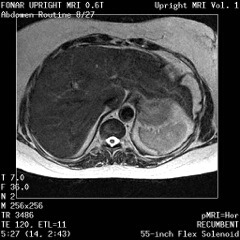 |
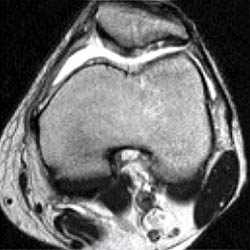
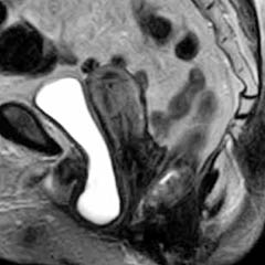
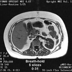
The Liver, Kidney and Small Intestine |
|
|
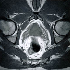
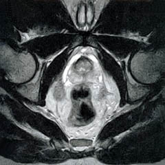
Bladder and Uterus in Pelvic Floor Dysfunction (PFD) |
|
|
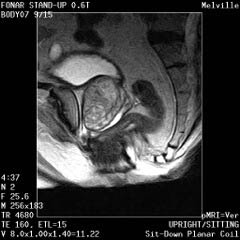
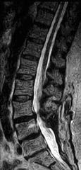
Lumbar Spine |
Recumbent, Weightless |
Upright, Weight-Bearing |
Figure 8.
Figure 8a-8d.
Further examples of the exceptional anatomic detail
made visible by the DISCOVERY
of Damadian of the pronounced differences in the decay
rates (relaxations) of the NMR signals
of the body's normal tissues (Figure
6). The DISCOVERED
differences supply the pixel amplitude differences
"PIXEL CONTRAST (IMAGE DETAIL)"
that produce, for the first time in medical history,
the detailed visualization of normal human anatomy
MRI is noted for. Note the visualization of the
vestibular and cochlear nerves
WITHIN
the internal auditory canel (Figure 8b) and the visualization
of the hypothalamic
tract (that transports hormones from
the brain) WITHIN
the pituitary stalk. (Figure 8c)
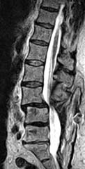
Lumbar Disc Herniation
Figure 24.
Merril Simon - Co-author
" The Pioneers of NMR and Magnetic Resonance
in Medicine - The Story of MRI", James Mattson
and Merrill Simon
Bar-Ilan University Press, 1969
On the Accomplishment
of the World's First MRI Scan
of the Live Human Body 7/3/1977
Figure 25.
Figure 26.
Figure 27.
Figue 28.
"We
are perplexed, disappointed and
angry about the uncomprehensible
exclusion of Professor Raymond Damadian M.D. from
this year's Nobel Prize in Physiology or Medicine.
MRI's entire development rests on the shoulders
of Damadian's discovery
of NMR proton relaxation differences among normal
and diseased tissues and his proposal of external
scanning of NMR relaxation differences in the
human body, published in Science in 1971"
Eugene Feigelson, M.D.
Dean of the College of Medicine
SUNY Downstate Medical Center
Distinguished Service Professor
Senior Vice President for Biomedical Education and Research
|
|
Figures
30a-30c. First ever MRI images of patients with cancer
(1978)
(obtained on Indomitable)
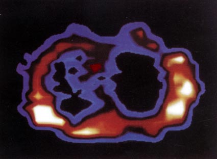 |
Figure 30a. FONAR scan at the level of 1-3/4 inches below the Angle of Lewis, by the method of Indomitable1, (Figs 20c and 20e) in a man 46 years old with pulmonary oat cell carcinoma. Tumour indicated by light blue infiltrate in left lung field, which should be black as it is in right lung cavity. Midline structure (red) separating the two lung cavities is the cross section through the arch of the aorta.
(
Philosophical Transactions of The Royal Society of London B, 1980, Vol. 289, pg 498, plate 2, figure 13.)
|
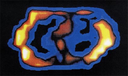 |
Figure 30b. FONAR cross-sectional scan through the thorax at the level of the 3rd intercostal space, by the method of Indomitable1, (Figs 20c and 20e) in a patient with an adenocarcinoma of the breast that metastasized to the right lung. The tumor is seen as a band of signal-producing tissue (light blue) bridging the right lung cavity. The tortuous structure separating the right and left lung cavities is the aortic arch. (1978) (Scanning time: 36 min.)
( Philosophical Transactions of The Royal Society of London B, 1980, Vol. 289, pg 497, plate 3, figure 15. (color version))
|
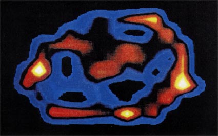 |
Figure 30c. FONAR cross-sectional scan through the low chest (10th thoracic vertebra), by the method of Indomitable1, (Figs 20c and 20e) in a patient with advanced alveolar cell carcinoma. The tumor is seen as intense signal-producing tissue (red, and less signal-intense light blue) invading both lung cavities and obliterating the bulk of the air space. (1978) (Scanning time: 30 min.)
( Philosophical Transactions of The Royal Society of London B, 1980, Vol. 289, pg 497, plate 3, figure 14. (color version))
|
1978
First Commercial MRI Company Founded
FONAR Corporation
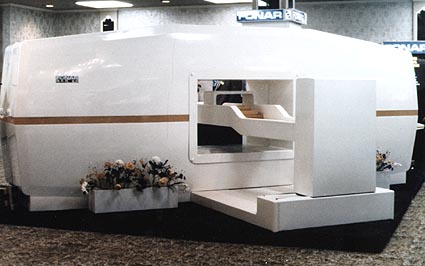 |
Figure 31. First-ever commercial MRI the FONAR QED 80. The FONAR QED 80 was equipped with a computer driven patient transport system (Vertical white bed mount pictured at the front of the QED 80 scanner) to automate the manual three-dimensional step-wise scanning procedure utilized by Indomitable1. (Indomitable transport apparatus Figs. 20c & 20e) |
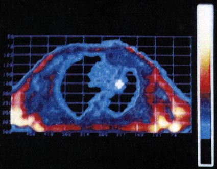 |
Figure 32. "Carcinoma of the left upper lobe with peripheral consolidation". Images published by Drs. Ross, Lie, Thompson & Associates from their FONAR QED 80 MRI scanner installed in their radiology practice in Clevelend, Ohio. QED 80 images were acquired by the step-wise 3D patient translocation method of Indomitable1.
(Radiology Nuclear Medicine Magazine, June 1981, cover) |
1. The 3D step-wise translocation of the patient across the magnetically focused resonance aperture. The resonance aperture was achieved by focussing the "near field" magnetic component of the transmitted rf (U.S. Patent 3,789,832) in combination with the shaping of the static magnetic field of the region of interest to generate a spatially localized NMR signal.
|
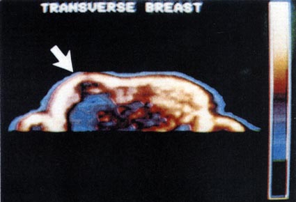 |
Figure 33. "NMR image of the breast shows a large mass (dark area) in the central portion of the right breast. T1 data are consistent with the diagnosis of cyst. (mean: 151, width: 239)" Images published by Drs. Ross, Lie, Thompson & Associates from their FONAR QED 80 MRI scanner installed in their radiology practice in Clevelend, Ohio. QED 80 images were acquired by the step-wise 3D patient translocation method of Indomitable1.
( Radiology Vol.143, No.1, pg 202, April 1982.)
|
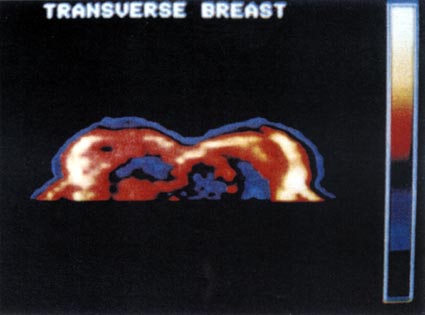 |
Figure 34. "NMR image shows a region of low density in the left breast with elevated T1 values. The small black area in the right breast is compatible with the diagnosis of cyst. (mean: 127, width: 128)" Images published by Drs. Ross, Lie, Thompson & Associates from their FONAR QED 80 MRI scanner installed in their radiology practice in Clevelend, Ohio. QED 80 images were acquired by the step-wise 3D patient translocation method of Indomitable1.
( Radiology Vol.143, No.1, pg 204, April 1982.)
|
The Bible teaches
“God hath made man Upright”
Ecclesiastes 7:29
So Why Not Scan Him the Way
He Was Made ?
2001
The Upright MRI Begins
Figure 35.
Figure 36.
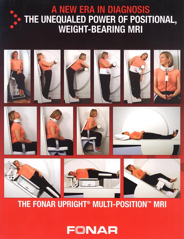
Figure 37.
The
Mommy and Me MRI
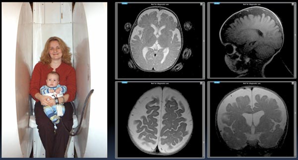
Figure 38.
The Mommy and
Me MRI
The Mommy and Me
MRI produced the above (just obtained) infant pictures
of a 7 month old child WITHOUT ANESTHESIA with
the infant lying in the scanner in a Fonar receiver
coil with the mother kneeling and facing into the scanner
(opposite to the position shown) holding the child's
head. The upper left image of the infant's brain shows
the mother's head positioning finger-tips. The brain
images obtained exhibit hydrocephalus in the infant
together with pronounced CSF pooling suggestive of significant
obstruction to the flow of CSF (most likely cervical
obstruction) in and out of the brain generating increases
in intracranial pressure (ICP) and cerebral pooling
of CSF as visualized in the above brain images of the
infant.
Figure 39.
THE TRUTH OF HISTORY, THE UNIVERSITY
OF CHICAGO PRESS – 2 YEARS AFTER THE NOBEL PRIZE
 “By
the final few decades of the twentieth century, medical
practitioners were exploiting developments in nuclear
physics to provide a range of new ways of peering
inside the human body …. Another technique developed
during the 1970s was MRI (magnetic resonance imaging).
The technique was initially developed by Raymond Damadian
(1936 -), working at the Downstate Medical Center
in New York, making use of the fact that different
atomic nuclei emit radio waves of predictable frequencies
when exposed to a magnetic field. Damadian
noted that tumorous cells emitted signalsdifferent
from those emitted by healthy tissue and used this
as the basis for a new technique for identifying cancers.
Damadian and his fellow workers produced the first
MRI scan of the human body in 1977.” “By
the final few decades of the twentieth century, medical
practitioners were exploiting developments in nuclear
physics to provide a range of new ways of peering
inside the human body …. Another technique developed
during the 1970s was MRI (magnetic resonance imaging).
The technique was initially developed by Raymond Damadian
(1936 -), working at the Downstate Medical Center
in New York, making use of the fact that different
atomic nuclei emit radio waves of predictable frequencies
when exposed to a magnetic field. Damadian
noted that tumorous cells emitted signalsdifferent
from those emitted by healthy tissue and used this
as the basis for a new technique for identifying cancers.
Damadian and his fellow workers produced the first
MRI scan of the human body in 1977.”
(Making Modern Science, A Historical
Review, The University of Chicago Press, 2005).
THE TRUTH OF
HISTORY, SUNY HEALTH SCIENCES CENTER (DOWNSTATE MEDICAL
CENTER), BROOKLYN – 5 YEARS AFTER THE NOBEL
PRIZE
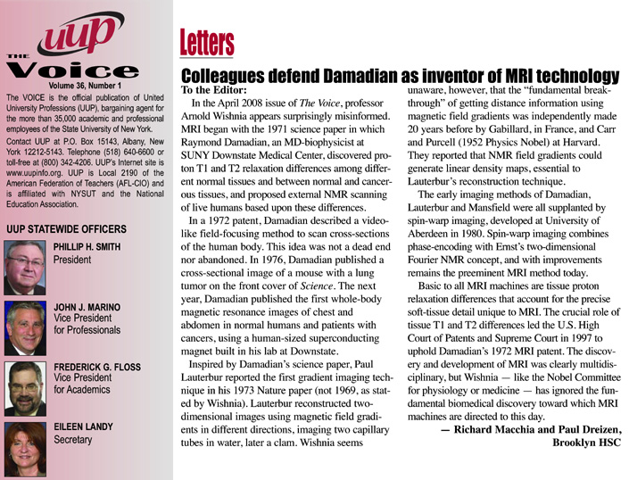
Figure 40.
(Richard Macchia, MD, and
Paul Dreizen, MD, in the UUP Voice, the official
publication of United University Professions (UUP),
State University of New York.)
American
Institute for Medical and Biological Engineering (AIMBE)
Washington, D.C.
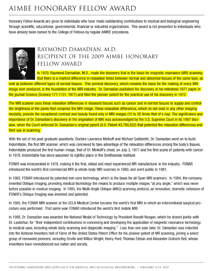 Figure 41.
Figure 42.
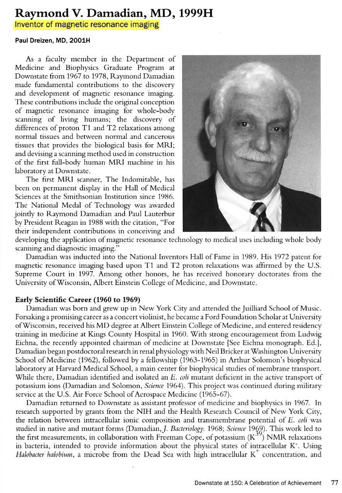
IN SUMMATION,
ORIGINATION
OF THE NMR BODY SCANNING IDEA
Damadian both
originated the IDEA
to scan the human body by NMR (MR) and provided
the means (the NMR
signal differences)
to achieve it. For any scientific advance to occur,
someone must generate the IDEA
to bring it about. Damadian
provided the IDEA
to give rise to the MRI.
Prior to Damadian's
genesis of the NMR body scanning IDEA
in 1969, (Figs. 1, 2, 3)
NMR instruments for obtaining the NMR spectra of
test tube samples had been in
operation for 23 years. Thousands of research
scientists and chemists the world over used NMR
spectrometers to obtain spectra of chemical samples,
(Fig 18) but the IDEA
to use NMR to scan the human body occurred
to no one.
For example,
without the IDEA
to create an independent self-contained transportable
COMBUSTION
ENGINE, there would be no AUTOMOBILE.
Without the IDEA
to transport vehicles by air flotation, there would
be no AIRPLANES.
Without the
IDEA
to generate light by electricity, there would be
no LIGHT
BULBS and without
the IDEA
to scan the human body by NMR
(Figs. 1, 2, 3)
there WOULD
BE NO MRI. The IDEA
to scan the HUMAN
BODY BY NMR ORIGINATED WITH DAMADIAN.
Indeed, when
Damadian first originated the IDEA
it was ridiculed at a major scientific conference
held at the National Cancer
Institute of the National Institutes of Health in
1976. A distinguished member of the NMR profession
and a professor at the Johns Hopkins University
at the time presented his successful repeat of Dr.
Damadian's NMR measurements of malignant tissue.
He went on to report that other non-malignant diseased
tissues also had elevated NMR relaxations. Overlooking
that both malignant tissues and non-malignant diseased
tissues had marked NMR relaxation elevations when
compared to normal that would generate marked differences
in pixel brightness (contrast) when displayed on
an MRI image he announced.
"Therefore any further discussion of scanning
the human body
by NMR is visionary nonsense." (Fig. 19b)
At another
NMR conference that Dr. Damadian was asked to present
at, the Moderator of the NMR conference stood up
after his presentation and asked
"Now Doctor, how fast do you propose to
spin the patient ?" (Fig. 19c)
The focused
rf of U.S. Patent 3,789,832 (Fig. 20c,
20e) together with shaping of
the static magnetic field provided the means for
the spatial localization needed for the in vivo
scanning of the 106 (Fig. 21
& Fig.22) anatomic loci
(pixels – see Fig. 10)
that provided the first MR image of the human body.
It was demonstrated at trial that the magnetic component
(the "near field" that generates the NMR
signal) of the
transmitted rf (radio frequency) could be shaped
to any dimension desired ("focused") and
moved to any location desired1.
The focusing of the transmitted rf would be used
to systematically generate the NMR signals
from spatially localized scanning loci within the
body, in order to map the anatomy and detect diseased
loci.
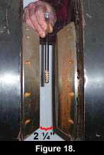 Except
for Damadian's BOLD
idea to take
the NMR test-tube analyzer and its 2 1/4 inch sample
space Except
for Damadian's BOLD
idea to take
the NMR test-tube analyzer and its 2 1/4 inch sample
space
(Fig 18) and totally remake the magnet and its millimeter
diameter NMR rf receiver coils*
that had been devoted unchanged for more than 23
years, (since the inception of NMR), to the chemical
analysis of test-tube samples, there would be no
MRI today.
Notwithstanding
grave doubts that there would be sufficient NMR
signal/noise
reception to detect a tiny NMR signal
from a small voxel (volume element) deep within
the interior of the human body, using a receiver
coil 10 inches away from the signal
generating volume, and still achieve an NMR signal
large enough to scan the entire human body, the
initiative for the NMR body scanner (MRI) proceeded
under the Good Lord's protection and direction (Col.
2:3).
It took Damadian
to envision the radically transforming step of taking
the traditional 23 year old NMR test-tube analyzer
and using it to invent a bold new revolutionary
scanner for the entire human body.
*rf
transmitter coils, rf receiver coils, pre-amplifiers,
rf tuned resonance circuits, etc that had been devoted
unchanged for more than 23 years since the inception
of NMR to the NMR chemical analysis of test-tube
samples.
1: see Fig.32
legend.
DISCOVERY
OF THE NMR SIGNAL
DIFFERENCES TO MAKE THE
IMAGE
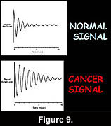 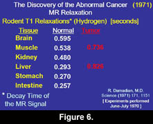 Having
originated the NMR body scanner IDEA,
Damadian also provided the means to bring it about.
He isolated the T1 and T2 tissue NMR relaxation
differences (See "T1 and T2 Imaging" section
above. Click here) that made
it possible. He demonstrated for the first time
that the NMR signal
from cancer tissue was markedly different from the
NMR signals obtained
from normal tissues. He further demonstrated in
the same 1971 publication in Science (Damadian,
R. Tumor Detection by Nuclear Magnetic Resonance.
Science, 171:1151-1153, 1971) that the NMR signals
of the normal tissues also differed markedly. Having
originated the NMR body scanner IDEA,
Damadian also provided the means to bring it about.
He isolated the T1 and T2 tissue NMR relaxation
differences (See "T1 and T2 Imaging" section
above. Click here) that made
it possible. He demonstrated for the first time
that the NMR signal
from cancer tissue was markedly different from the
NMR signals obtained
from normal tissues. He further demonstrated in
the same 1971 publication in Science (Damadian,
R. Tumor Detection by Nuclear Magnetic Resonance.
Science, 171:1151-1153, 1971) that the NMR signals
of the normal tissues also differed markedly.
|
Figure 14 is an axial
(cross-sectional) image of the brain showing
a tumor of the cerebellum (white areas)
in the midline. Figure 14c is a magnified
image showing the picture elements or "pixels"
(small squares) that make up the image.
The cerebellar
tumor as it would appear (D) with no MR signal
differences. Figure D is the same image as
B but where all MR signal
differences were eliminated and all the MR
pixels therefore had the same pixel brightness.
The absence of the MR signal
differences between cancer and normal tissue
discovered
by Damadian gives the MR image pixels equal
brightness and the
tumor becomes Invisible. |
Were the amplitudes
of the NMR signals
(fig.9) used to set the brightness
of each MRI image pixel the same for all tissues
(and prior to Dr. Damadian's discovery
such NMR tissue signal
differences were not known to exist) the brightness
of each image pixel would be the same. The MR image
would be a blank.
The NMR signal
differences discovered
by Damadian (Figs 6,9,tables
1 & 2) vary the brightness
of the pixels that make up the image (Figs. 6,9).
The signal differences
of diseased and normal tissues generate the large
differences in pixel brightness that enable all
diseased tissues (cancerous as well as non-cancerous)
to be exquisitely visualized (fig.3b,10,11-13)
by the MRI image. Additionally the exceptional NMR
signal differences
among the normal tissues discovered by Damadian
give rise to the extraordinary detail of normal
anatomy visualized by MRI (figs. 7,8)
Except for the
marked NMR signal
differences between diseased tissues and normal
tissues discovered
by Damadian and the marked differences of the NMR
signals within
the normal tissues themselves discovered
by Damadian there would be no MRI. It is the tissue
NMR signal differences
discovered by
Damadian that make the MRI image.
The tissue MR (NMR) signal
differences discovered
by Damadian are the BUILDING BLOCKS from which the
MRI IMAGE is CONSTRUCTED.
Without the BUILDING BLOCKS there is no BUILDING!
Without the tissue NMR signal
differences needed to construct an image, that were
discovered by Damadian
(and EXCLUDED by the NP),
THERE IS NOTHING TO MAKE THE IMAGE WITH
and
THERE IS NO MRI!
Except
for the NMR signal
differences discovered
by Damadian to
MAKE THE IMAGE
THERE IS NO IMAGE !
If
the NMR signals
used to set the brightness of the image
pixels do not vary from diseased to normal
tissues or within the normal tissues themselves,
as discovered
by Damadian*, then
THERE IS NOTHING TO MAKE THE MRI IMAGE WITH!
The image pixels that the MR scanner
MAKES THE IMAGE WITH
will all have the same brightness and
THE MRI IMAGE WILL BE A BLANK !
|
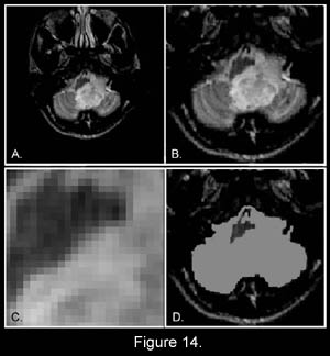
Figure 14 is an axial (cross-sectional) image of the brain showing a tumor of the cerebellum (white areas) in the midline. Figure 14c is a magnified image showing the picture elements or "pixels" (small squares) that make up the image.
The
cerebellar tumor as it would appear (D)
with no MR signal
differences. Figure D is the same image
as B but where all MR signal
differences were eliminated and all the
MR pixels therefore had the same pixel brightness.
The absence of the MR signal
differences between cancer and normal tissue
discovered
by Damadian gives the MR image pixels equal
brightness and the tumor becomes Invisible.
|
EXCLUDED
INTENTIONALLY
Figure 19 Nobel Statutes
"If a work which is to be rewarded has been produced by two or three persons, the prize shall be awarded to them JOINTLY. In no case may the prize be divided between more than THREE PERSONS." (Nobel Statutes)
The 2003 Nobel Prize for MRI, however was awarded to two persons. P.C. Lauterbur and P. Mansfield, not to the three persons provided for by the Nobel Statutes. The Third Nobel award provided for, and used many times throughout the history of the Nobel Prize, was intentionally VOIDED.
Dr. Damadian was thereby EXCLUDED by the NOBEL (and therefore from history) from any recognition for his critical role in the genesis of the MRI without which MRI WOULD NOT EXIST !
The Nobel Statutes
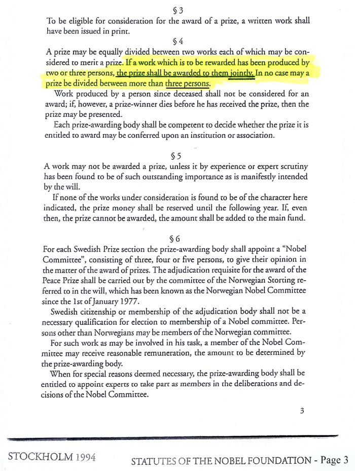
Thus Dr. Damadian's absence from the Nobel Prize for MRI was not an oversight. He was
EXCLUDED INTENTIONALLY
Remarkably, EXCLUDING Dr. Damadian
constitutes EXCLUDING
THE ONE ORIGINATOR WITHOUT WHOM THERE IS NO
MRI AT ALL !! It constitutes EXCLUDING
the person who for the
first time ever in history conceived
of the idea
to use a 23 year old test-tube analyzer (Fig.
18, the NMR test-tube analyzer) to scan the
live human body, an idea
branded as "visionary nonsense" at
the time it was originated (Fig. 19b). It also
constitutes EXCLUDING the person who further,
for the first time ever,
PROVIDED the MEANS to ACHIEVE AN MRI IMAGE.
HE PROVIDED THE SIGNAL TO
MAKE THE IMAGE WITH (Figs. 4a, Tables I &
II, Fig. 4b, Fig. 6) and WITHOUT
WHICH THERE IS NO MRI IMAGE AT ALL !!
It is indisputable that Dr. Damadian is the vital contributor by whom (in accordance with the Nobel Statutes) "the work which is to be awarded has been produced."
Indeed as Professor
Henry Wallman, Professor of Mathematics, Massachusetts
Institiute of Technology (MIT) stated,
"I
am of the definite opinion that Dr. Damadian's
contribution was both prior to and more
fundamental than Dr. Lauterbur's"
(underlining included in original [ J. Mattson
and M. Simon "The
Pioneers of NMR and Magnetic Resonance in Medicine:
The Story of MRI" Bar-Ilan University Press
(1996), 676 ].
Notwithstanding, Dr. Damadian's
vital role in the genesis of MRI and the specific
provision of the Nobel Statues to provide the
award to up to "THREE
PERSONS"
"JOINTLY",
the Nobel Committee intentionally subverted
this option (of the Nobel Statutes) and EXCLUDED
Dr. Damadian. Remarkably in VOIDING
the third award provided by the Nobel Statutes
the Nobel Committee intentionally
and untruthfully EXCLUDED the only
MRI innovator without whom THERE
IS NO MRI AT ALL! They EXCLUDED the innovator
who originated the idea
(Fig. 17d) of the MR body scanner for medical
disease and who proposed FOR
THE FIRST TIME EVER that the 23 year
old non-invasive NMR analyzer of test-tube samples
could be used to scan the live human body to
detect disease and without which idea
no such scanner could ever come into existence.
They also EXCLUDED the only one who DISCOVERED
the NMR SIGNAL that could achieve it, the NMR
SIGNAL that makes the image and without which
THERE IS NO IMAGE!
VIOLATION OF THE WILL OF
ALFRED NOBEL
For his prize in physics, Nobel specifies it is to be given for the "most important "discovery OR invention". For his prize in chemistry, Nobel specifies it is to be given for the "most important chemical discovery OR improvement".
In so doing, Nobel defines what a discovery is according to HIS WILL.
It is NOT a method !
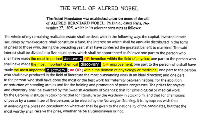
DELIBERATE VIOLATION
OF
ALFRED
NOBEL'S WILL !
the 2003
Nobel Committee
REMARKABLY
EXCLUDED
the only
GENUINE
DISCOVERY
that
QUALIFIED
for
the NOBEL PRIZE
in MEDICINE
under Alfred Nobel's WILL,
Dr. Damadian's DISCOVERY
of the
abnormal NMR signals of
diseased tissue
THAT
MAKE THE IMAGE,
while granting the NOBEL AWARD
INSTEAD to two (METHODS)
that
DID
NOT QUALIFY
for the Nobel Prize in
MEDICINE under Alfred Nobel's WILL1
1
The innovations of METHODS
that QUALIFY
for the NOBEL PRIZE in Physics and in Chemistry
under Alfred Nobel's WILL
were explicitly EXCLUDED
by Nobel in his WILL
for his AWARD in MEDICINE
which states that his award
in medicine is to be given for
DISCOVERY
ONLY.
As
Nobel specifies in his WILL
his AWARD
in MEDICINE shall be given
"to
the person who shall have made the most important
DISCOVERY
(no OR)
within
the domain of physiology or MEDICINE "
(ONLY)
in
contrast to Nobel's award in physics which states
"to
the person who
shall have made the most important
DISCOVERY
OR
Invention
within
the field of physics"
and
in contrast to Nobel's award in chemistry which
states
"to
the person who
shall have made the most important chemical
DISCOVERY
OR
Improvement
".
IN
ORDER TO CIRCUMVENT
NOBELS's
EXCLUSION of METHODS
from the NOBEL
PRIZE in MEDICINE
the 2003 NOBEL Committee
FALSELY
DESIGNATED
the Lauterbur and Mansfield
contributions as "discoveries"
(i.e.
their Nobel citation -
"for their discoveries
concerning magnetic resonance imaging")
when they were
not "discoveries"
at all, but
were EXCLUSIVELY
METHODS1,2
(and ONLY
METHODS)
that
DID NOT QUALIFY
for the Nobel Prize in Medicine under Alfred
Nobel's WILL.
They were not
GENUINE DISCOVERIES
of a new scientific phenomenon
such as
Dr. Damadian's
GENUINE
NEW SCIENTIFIC DISCOVERY
,for
the first time ever,
of the
PREVIOUSLY UNKNOWN
ABNORMAL NMR SIGNALS
of
DISEASED TISSUE
(and
their large variations among healthy tissues)
that are used to
GENERATE ALL OF TODAY'S
MRI IMAGES
(and
WITHOUT WHICHTHERE
WOULD BE NO MRI IMAGE AT ALL
!)
They
were, instead,
EXCLUSIVELY
METHODS
2
created to generate
pictures of the abnormal NMR signals of diseased
tissue
GENUINELY
DISCOVERED
by
Dr. Damadian
which SIGNALS
ARE USED
TO MAKE
all of
TODAY'S
MRI IMAGES !
THUS,
in SUMMATION
the
2003 Nobel Committee
SPECIFICALLY
EXCLUDED the
only innovation
that
QUALIFIED
for
the Nobel Prize
in Medicine under Alfred
Nobel's WILL while they made the award
instead to
method innovations 2
EXPLICITLY
EXCLUDED
by Alfred Nobel from the
Nobel Prize in Medicine !
2
Lauterbur provided the METHOD
of using a series of magnetic field gradients
to generate a picture of the abnormal NMR signals
of diseased tissues DISCOVERED
by Damadian.
Mansfield provided ONE
of the METHODS
of making a picture from the spin echo of the
NMR signal, the Echo Planar Image, of the abnormal
NMR signals of diseased tissues DISCOVERED
by Damadian.
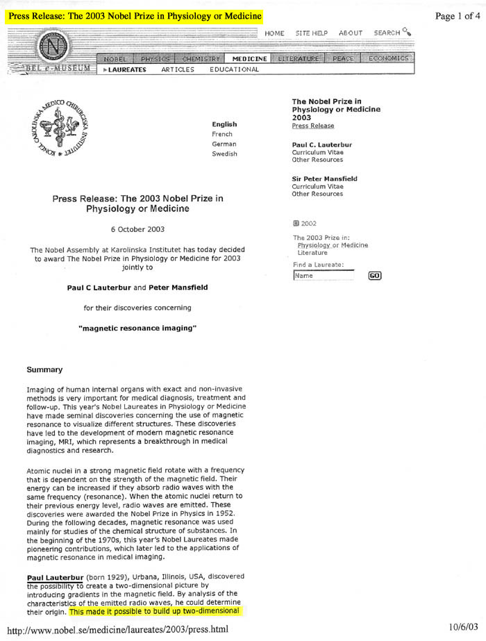
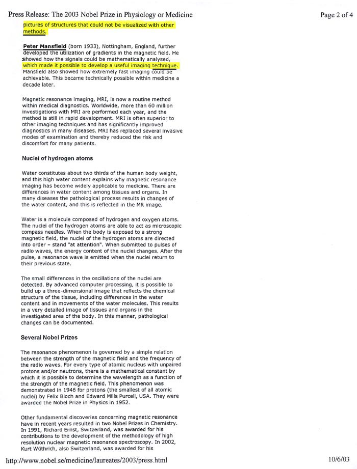
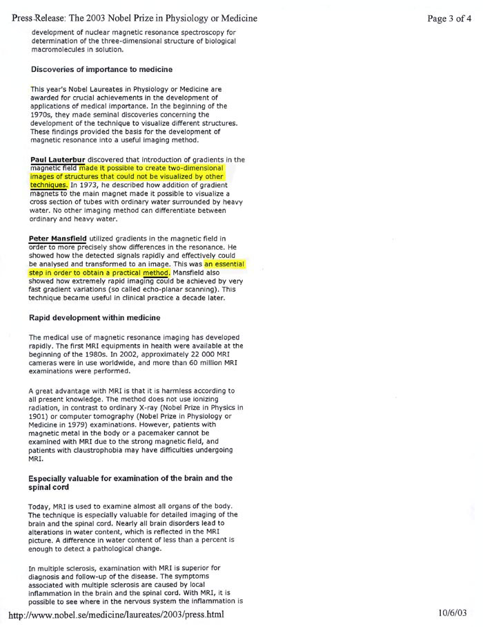
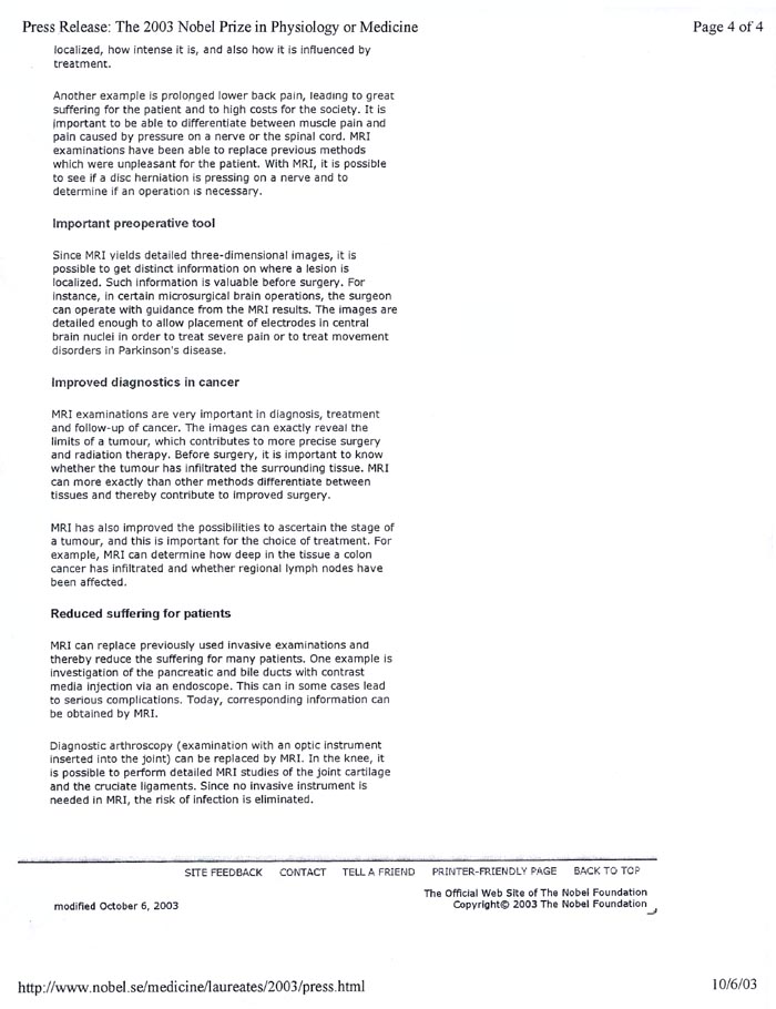
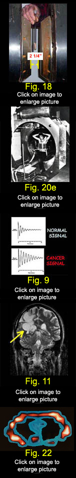 Apart from Dr. Damadian's
ORIGINATION
of the
IDEA*
to take a 23 year old
2¼ inch test-tube analyzer (Fig 18) and
REDESIGN AND TRANSFORM IT into a non-invasive scanner
for detecting disease in the live human body (Figs
1,2,3,20e)
and
Apart from Dr. Damadian's
DISCOVERY**
of the
disease detecting signal
to do it with
(Figs 3,4a,4b,9,11: tables 1 & 2)
THERE WOULD BE NO MRI TODAY !!
(Fig 22, Fig 11)
"About 22,000 MRI machines around the world were used in 60 million examinations last year"
(The Economist, Dec. 4, 2003 (Q4 2003))
* An
IDEA
publically branded "NONSENSE"
at the time (Cursor click on
the black rectangle of interest to visit the cited
reference)
**
... the
discovery that
tissue disease altered the NMR radio signal produced
by the body's tissues (Figure
9). The diseased tissue NMR radio signals
obtained in an NMR scan of the live human body
were thereby made distinct
(Figure 9) from
the tissue NMR radio signals obtained from the
bodies surrounding normal tissues (Figure
11) . The discovery
enabled detailed visualization of the bodies'
diseased tissues (Figure
11) for the first time in medical
history ever thereby making possible NMR (MRI)
scans of the live human body (enabling
early treatment).
In so far as
Without Edison there is no Light Bulb
Without Alexander Graham Bell there is no Telephone
Without the Wright Brothers there is no Airplane
Without Damadian there is no MRI*
*as affirmed by
TWO PRESIDENTS OF THE UNITED STATES,
THE NATIONAL INVENTORS HALL OF FAME,
THE UNITED STATES SUPREME COURT.
"but in the end
THE TRUTH WILL OUT"
William Shakespeare (1598 AD)
"The Merchant of Venice"
Launcelot Gobbo to Old Gobbo (Launcelot's father)
i.e. despite
the
Nobel Committee's effort
to
erase Dr. Damadian
from the history of his invention and from his
7 years
of suffering to surmount the
"INDOMITABLE"
technological obstacles
to convert
a 10 mm test-tube analyser
into a scanner of the LIVE human body
what the Nobel Committee appears not to appreciate, in contrast to
Launcelot Gobbo,
is that
THE TRUTH
IS
INVINCIBLE !!
|