Images courtesy of Imaging Center At
Boot Ranch
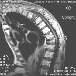
Severe
Kyphosis Rendering Recumbent Imaging Impossible
Sagittal images of the lumbosacral (6A) and thoracic (6B)
spines in the upright-seated position shows compression of
two thoracic vertebral bodies. This was ultimately found to
be due to osteoporosis. The patient suffered from sufficiently
marked kyphosis to render recumbent imaging impossible by
either computed tomography or MRI. |