|
Recumbent
|
Standing Extension
|
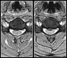
Recumbent
|
Standing Extension
|
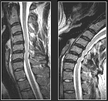  Images courtesy of Melville MRI, P.C
Case Study:
Upright Dynamic MRI Reveals Hidden
Disc Herniation
The axial standing-extension gradient echo image (right) demonstrates
a focal posterior disc herniation at the C4/5 level not visible
on the recumbent scan. Note the associated spinal cord compression
on the standing-extension scan. |
Site Map
| Terms of Use-Our
Privacy Policy Use
Copyright © 2003 FONAR- All Rights Reserved |
