|
Severe Spondylolisthesis Undetected
by Recumbent MRI
Clinical Case Overview
A 57-year old woman presented with pain of one year's duration following
failed back surgery performed in 2001*.
The patient continued to experience persistent low back-pain,
accompanied by sensations of coldness and numbness in both thighs
and legs. The patient often required mechanical support to stabilize
her walking.
During the year following surgery, the patient sought help from
multiple medical specialists. She provided her recumbent MRI images
to them. She was told the images showed nothing that could account
for her symptoms and that nothing more could be done. Her surgeon
rejected the prospect of additional surgery. A Florida neurologist
suggested to her that her problem was “in her head.”
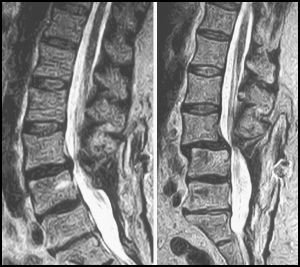
| Recumbent |
Upright |
 |
The imaging center that evaluated her recommended she be scanned in
an Upright™ MRI due to the possibility that an Upright™
scan, unlike the conventional recumbent scan, is weight-bearing and
"might uncover something." Her family physician wrote the
prescription, and the patient drove from her home in the Florida panhandle
to the closest FONAR Upright™ MRI center, which at the time
was in Tampa over 425 miles away.
The patient was scanned in the patented FONAR Upright™ MRI
in early 2002, one year after her spinal fusion. Both Upright™
and recumbent scans were performed on her in the multi-position
FONAR Upright™ MRI.
The recumbent MRI (left image) exhibited only a normal lumbar
lordotic curve and a modest bulge of the L3-4 intervertebral disc,
consistent with her prior recumbent MRI scans. The FONAR Upright™
scan (right image) revealed, however, a marked position-dependent
subluxation (anterolisthesis) at L3-4 and an accompanying spinal
stenosis that were not visible on the recumbent MRI.
The patient's Upright™ images established that there was
a genuine physical basis for her symptoms, whereas her recumbent
MRI images had failed to do so. The new Upright™ images supplied
her surgeon with the necessary evidence that additional surgery
was warranted to correct her problem.
A spinal fusion was performed at L3-4 one month after the patient's
Upright™ MRI scan. The surgical outcome was positive. To date,
almost four years post-op, the patient remains symptom free and
reported to FONAR, "Thank you for giving me my life back."
* laminectomy and L45S1 fusion

Manuel S. Rose, M.D.
Radiologist
Rose Radiology Centers
Florida, USA

Site Map
| Terms of Use-Our
Privacy Policy Use
Copyright © 2003-2006 FONAR- All Rights Reserved |
